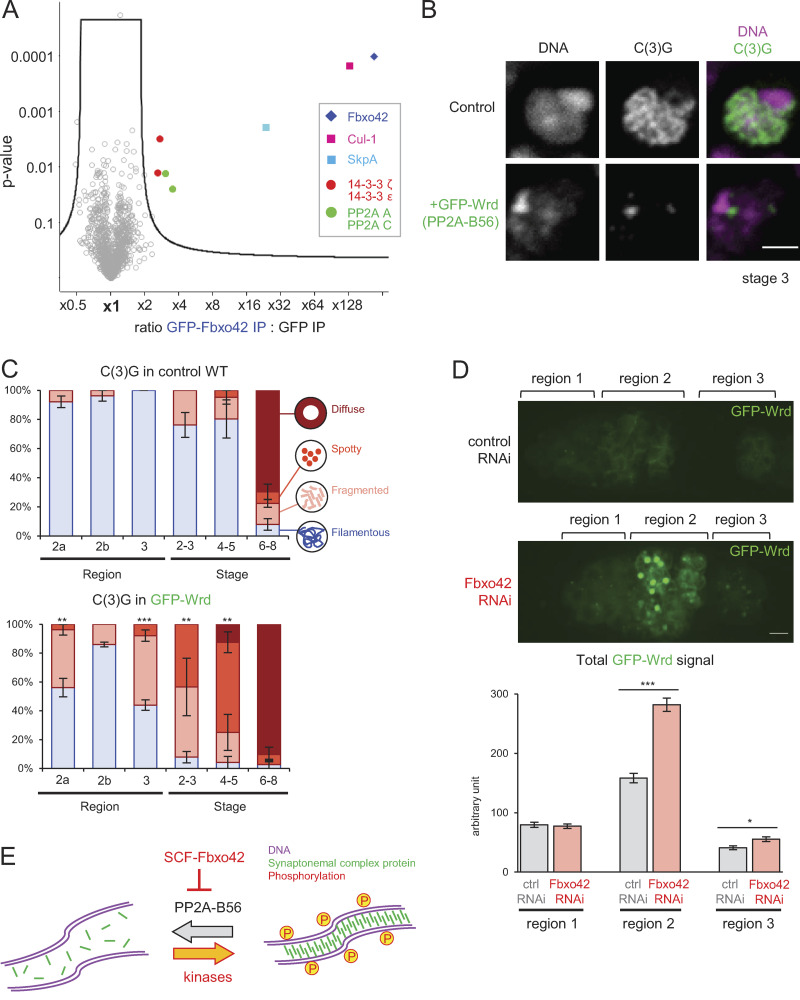
Figure 4.
Fbxo42 depletion increases PP2A-B56 (Wrd) levels, which destabilize the synaptonemal complex. (A) A volcano plot of proteins immunoprecipitated with GFP-Fbxo42 or GFP from ovaries. For each protein, the mean ratio of signal intensities found in immunoprecipitates of GFP-Fbxo42 and GFP alone was plotted against the statistical significance (the P value given by t test) from biological triplicates. SCF subunits, PP2A core subunits and 14–3-3 proteins were significantly enriched in GFP-Fbxo42 immunoprecipitates. (B) Localization of the synaptonemal complex component C(3)G in meiotic cells expressing GFP-Wrd (PP2A-B56) and a control. Scale bar = 3 µm. GFP-Wrd, nos-GAL4(MVD1) Wrd+/UASp-GFP-Wrd Wrd+; control, nos-GAL4(MVD1) Wrd+/Wrd+. (C) C(3)G localization patterns in meiotic cells overexpressing GFP-Wrd (PP2A-B56) and control cells throughout meiotic progression. Error bars represent SEM from triplicated experiments representing 19–24 germaria/oocytes for each region/stage. (D) GFP-Wrd signals in control and Fbxo42 RNAi live germaria. The images were captured and processed using identical conditions to allow accurate comparison. Total GFP signals above the background signals were quantified in regions 1, 2, and 3 of germaria (zygotene to early pachytene) expressing GFP-Wrd with control or Fbxo42 shRNA. Error bars represent SEM from analysis of 32–54 germaria for each. Scale bar = 2 µm. Fbox42 RNAi, nos-GAL4(MVD1) Wrd+/UASp-GFP-Wrd Wrd+ UASp-shRNA(FBxo42); control RNAi, nos-GAL4(MVD1) Wrd+/UASp-GFP-Wrd Wrd+ UASp-shRNA(white). (E) A schematic model showing that SCF-Fbxo42 down-regulates PP2A-B56 to tip the balance of phosphorylation toward synaptonemal complex assembly. *, P < 0.05; **, P < 0.01; ***, P < 0.001.

