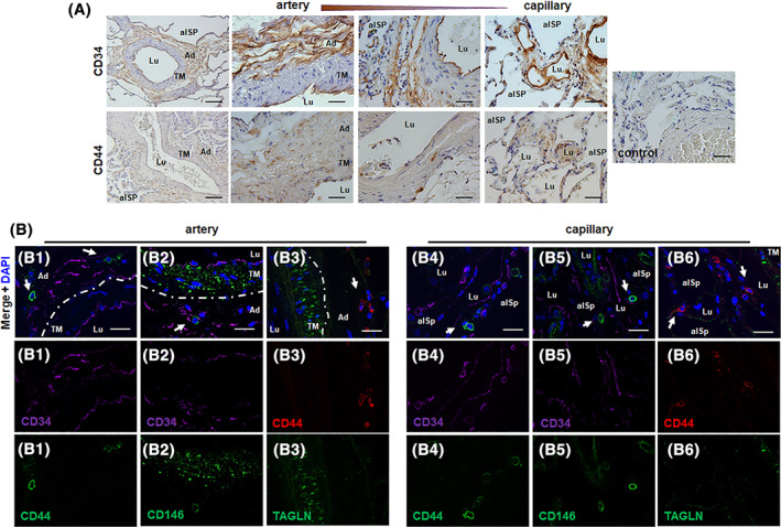
FIGURE 1.

The “vasculogenic zone” of lung blood vessels and co‐localization of mesenchymal stem cell (MSC) marker proteins (putative MSCs). A, Immunohistochemical staining of normal lung tissue sections was performed using antibodies against CD34 (EPC, endothelial progenitor cell marker) and CD44 (MSC marker) and DAB staining (brown). Representative lung photographs of larger blood vessels (with a thick muscular wall, >500 μm external diameter), in intermediate/small‐sized arteries (with thin muscular wall, up to 100 μm in external diameter) and in the microvessels (capillaries) are shown. Nuclei were counterstained with Hemalaun (blue). Lu lumen, TM tunica media, Ad adventitia, alSp alveolar space. Scale bar indicates 100 μm (left panel) and 10 μm (higher magnification images). B, Double‐immunofluorescent staining of normal lung sections was performed using antibodies against the typical MSC maker proteins CD44 (B1, B4) and CD146 (B2, B5) together with the endothelial and hematopoietic progenitor cell marker CD34, and of CD44 together with the smooth muscle cell marker transgelin (TAGLN, B3, B6). The dashes line emphasizes the border between the smooth muscle cell layer (TM) and the adventitia. Nuclei were visualized using DAPI nuclear counterstainings (blue). Representative lung photographs of arteries and microvessels (capillaries) are shown. Scale bar indicates 10 μm
