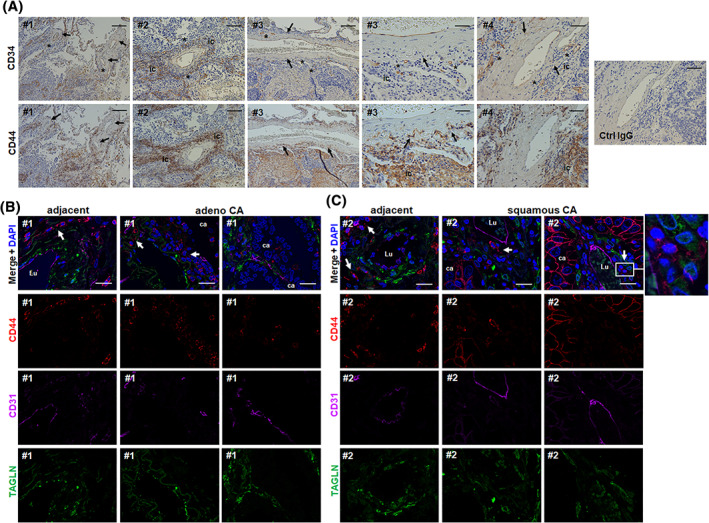
FIGURE 4.

The “vasculogenic zone” of lung blood vessels in NSCLC tissues and co‐localization of CD44‐positive vascular stem cells (LR‐MSCs). A, Immunohistochemical staining of paraffin‐embedded human NSCLC tissue sections were performed using antibodies against CD34 and CD44 in combination with DAB staining (brown). Representative photographs of large and intermediate‐sized lung blood vessels are shown. Arrows point toward cells residing in the vasculogenic zone. Asterisks highlight the vanishing of CD34 immunoreactive cells indicating that endothelial progenitor cells mobilize out of that zone (toward the tumor). Nuclei were counterstained with Hemalaun (blue). # numbers indicate NSLC tissues from different patients. ic, infiltrating immune cells. Scale bar indicates 100 μm (left panels) and 20 μm for the higher magnification images (right panel). B and C, Triple‐immunofluorescent staining of NSCLC specimen were performed using antibodies against the typical MSC maker protein CD44 (red) together with the smooth muscle cell marker transgelin (TAGLN, green) and the EPC marker CD34 (purple). Nuclei were visualized using DAPI (blue). Representative lung photographs of larger tumor blood vessels as found in adenocarcinoma (adeno‐CA) (B) and squamous cell carcinoma (squamous CA) (C) as well as of arteries present in adjacent normal lung tissue are shown. Different regions (blood vessels) of the same donor (#) are presented. Arrows point toward CD44‐positive cells residing in the vasculogenic zone. Lu, vessel lumen; ca, carcinoma cells. Scale bar indicates 10 μm
