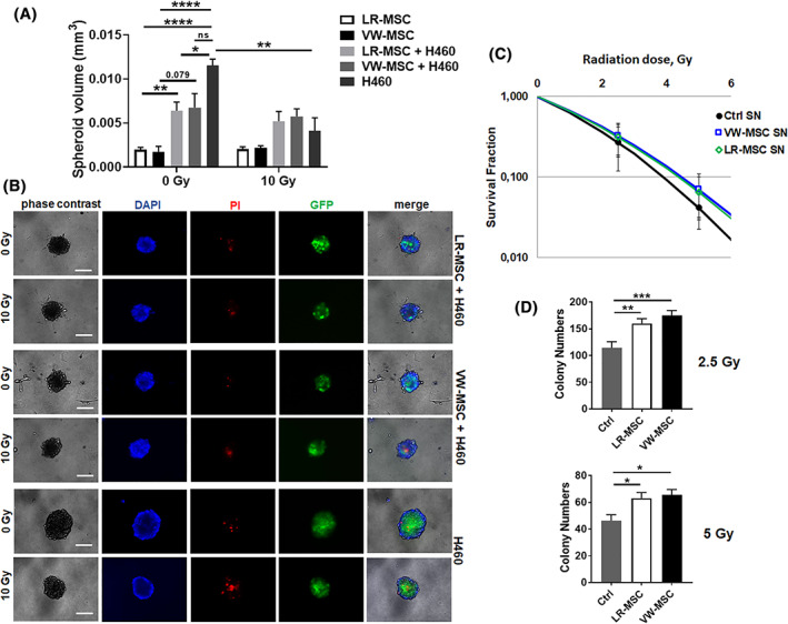
FIGURE 6.

Interaction of LR‐ and VW‐MSCs with lung cancer cells. A, NCI‐H460 lung cancer cells labeled with green‐fluorescent protein (GFP) were cultured alone or co‐cultured with LR‐MSCs or VW‐MSCs in hanging drops for 24 hours. After formation of spheroids, cells were plated in GFR‐Matrigel mixed with normal growth medium (1:2, vol/vol) and left untreated or irradiated at 10 Gy. Spheroids growth were measured after additional 48 hours of cultivation and respective volumes were calculated. Graphs depict the measurements from at least three independent experiments (H460, n = 3, LR‐MSC and LR‐MSC + H460, n = 6, VW‐MSC and VW‐MSC + H460, n = 5) where at least 10 spheroids per condition each were measured. *P < .05, **P < .01, ****P < .001 by one‐way ANOVA followed by post hoc Dunnett's test. B, Cell death was analyzed afterwards (48 hours' time point) by fluorescence microscopy using propidium iodide. DAPI was used for nuclei staining. Representative phase contrast images and simultaneously recorded fluorescent photographs from three individual experiments are shown (48 hours' time point). Scale bar represents 25 μm. C, Lung cancer cells were plated at low densities (CFU assay), irradiated with indicated doses (0, 2.5, 5 Gy) and further incubated for additional 10 days in conditioned media (SN) derived from cultured LR‐ and VW‐MSCs. Quantification of grown/surviving colonies was performed after Coomassie Brilliant Blue staining (Ctrl SN, n = 3, LR‐MSC SN, n = 6, VW‐MSC SN, n = 5). P by two‐way ANOVA, followed by post hoc Dunnett's multiple comparisons test: *P ≤ .05, **P ≤ .01, ***P ≤ .005
