
Figure 2. Activation of p38MAPK/ATF-7 in Intestinal Cells Suppresses Rotenone-induced Degeneration of Dopaminergic Neurons.
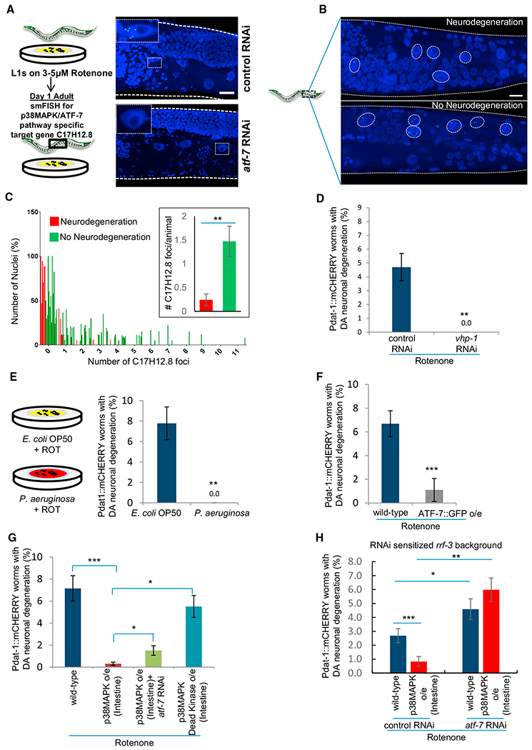
(A) Representative maximum projection confocal images of the ATF-7 target gene C17H12.8 mRNA in adult day 1, rotenone-treated animals on control and atf-7 RNAi. mRNA was detected by single-molecule RNA in situ hybridization (smFISH). Scale bar, 10 μm. n = 3 animals each treatment. Inset: single intestinal nucleus.
(B) Representative maximum projection confocal images of C17H12.8 mRNA in rotenone-treated animals that did, and did not, undergo neurodegeneration. Circles represent intestinal nuclei. Scale bar, 10 μm.
(C) Frequency distribution of percent of intestinal nuclei (y axis) that expressed 0–11 discrete C17H12.8 mRNA foci/nucleus (x axis) and mean number of C17H12.8 mRNA foci/animal counted in ~15 intestinal nuclei/animal (inset). Rotenone-treated wild-type animals that lost (red), and did not lose (green) dopaminergic neurons. n = 88–215 nuclei, from 6 to 16 animals.
(D) Percentage of animals subjected to vhp-1 RNAi that lost dopaminergic neurons on rotenone compared to control. n = 200–250 animals.
(E) Percentage of wild-type animals grown on Pseudomonas aeruginosa (PA14) that lost dopaminergic neurons on rotenone compared to control (OP50). n = 400 animals.
(F) Percentage of animals overexpressing ATF-7 that lost dopaminergic neurons on rotenone compared to wild-type. n = 300 animals.
(G) Percentage of wild-type animals, animals overexpressing p38MAPK in intestinal cells on control and atf-7 RNAi, and animals overexpressing kinase-dead p38MAPK in intestinal cells that lost dopaminergic neurons on rotenone treatment. n = 1,200 and 500 for wild-type animals and animals overexpressing active p38MAPK respectively and 127 for animals overexpressing intestinal kinase-dead p38MAPK.
(H) Percentage of rrf-3(mg373) animals and rrf-3(mg373) animals overexpressing p38MAPK in intestinal cells on control and atf-7 RNAi that lost dopaminergic neurons on rotenone. Data from paired experiments and controls are compared.
(C–H) Bar graphs show mean ± SEM. *p < 0.05, **p < 0.01, ***p < 0.001 (Student’s t test). Animals: day 1–2 adults. See also Figure S2.