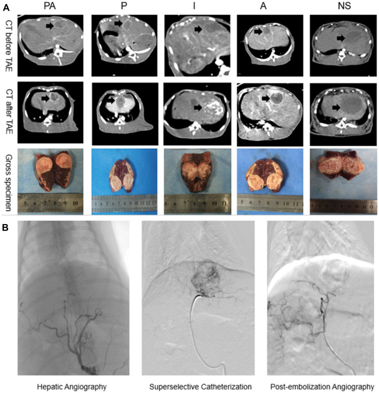
Figure 1.
(A) Abdominal contrast-enhanced CT scan was performed on VX2 tumor-bearing rabbits before and after TAE. We could see the nearly-circular tumor in the left lobe of liver (black arrows). (B) Hepatic artery angiography of rabbit liver before embolization, then super-selective catheterization and administer by the group. Blood supply of tumor was occluded and reconfirmed by post-embolization angiography.

