Figure 7.

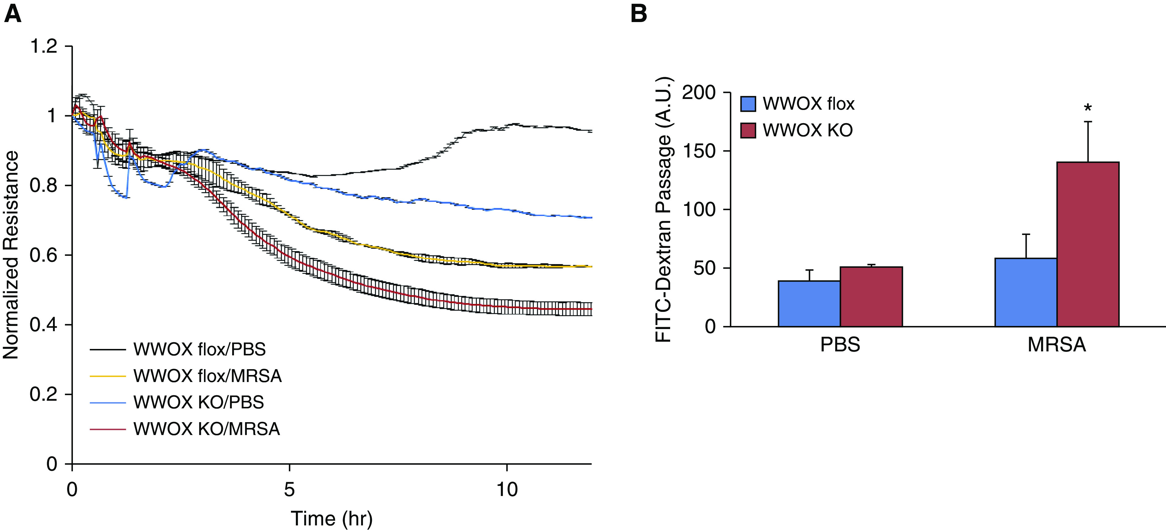
ECs isolated from EC WWOX KO mice are more susceptible to MRSA-induced monolayer barrier disruption. ECs from WWOX KO and WWOX flox controls were isolated by flow cytometric sorting for CD31+ CD45− cells and grown in culture. They were then examined by ECIS measurement of transendothelial resistance (TER) during exposure to heat-killed MRSA. (A) The scatter plot depicts changes in TER over the course of 10 hours following introduction of MRSA to the cell culture media. (B) In separate experiments, ECs from WWOX KO and WWOX flox controls were grown in a monolayer on transwell inserts containing 3-micron pores. Then, 10 μg/ml FITC-labeled dextran (3 kD) was added over the cells along with MRSA. After 3 hours, media from below the transwell was analyzed by fluorometry for the presence of FITC. Results are shown as means ± SD in n = 3 independent experiments. *P < 0.05, compared with MRSA-treated control by Student’s t test. A.U. = arbitrary unit.
