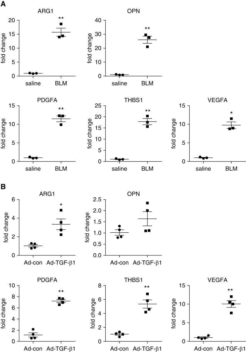
Figure 2.
Alveolar macrophages in fibrotic lungs demonstrate profibrotic phenotype. (A) C57BL/6 mice were i.t. instilled with saline or BLM. Three weeks after treatment, BALFs were harvested, and alveolar macrophages were isolated. Total RNAs were purified, and concentrations of the indicated genes were determined by real-time PCR (n = 3 mice for each group; mean ± SEM). *P < 0.05 and **P < 0.01 by two-tailed Student’s t test. (B) C57BL/6 mice were i.t. instilled with Ad-con or Ad-TGF-β1. Two weeks after treatment, alveolar macrophages were isolated, and total RNAs were purified. Concentrations of the indicated genes were determined by real-time PCR (n = 4 mice for each group; mean ± SEM). *P < 0.05 and **P < 0.01 by two-tailed Student’s t test. ARG1 = arginase 1; OPN = osteopontin; PDGFA = platelet-derived growth factor A; THBS1 = thrombospondin 1; VEGFA = vascular endothelial growth factor A.

