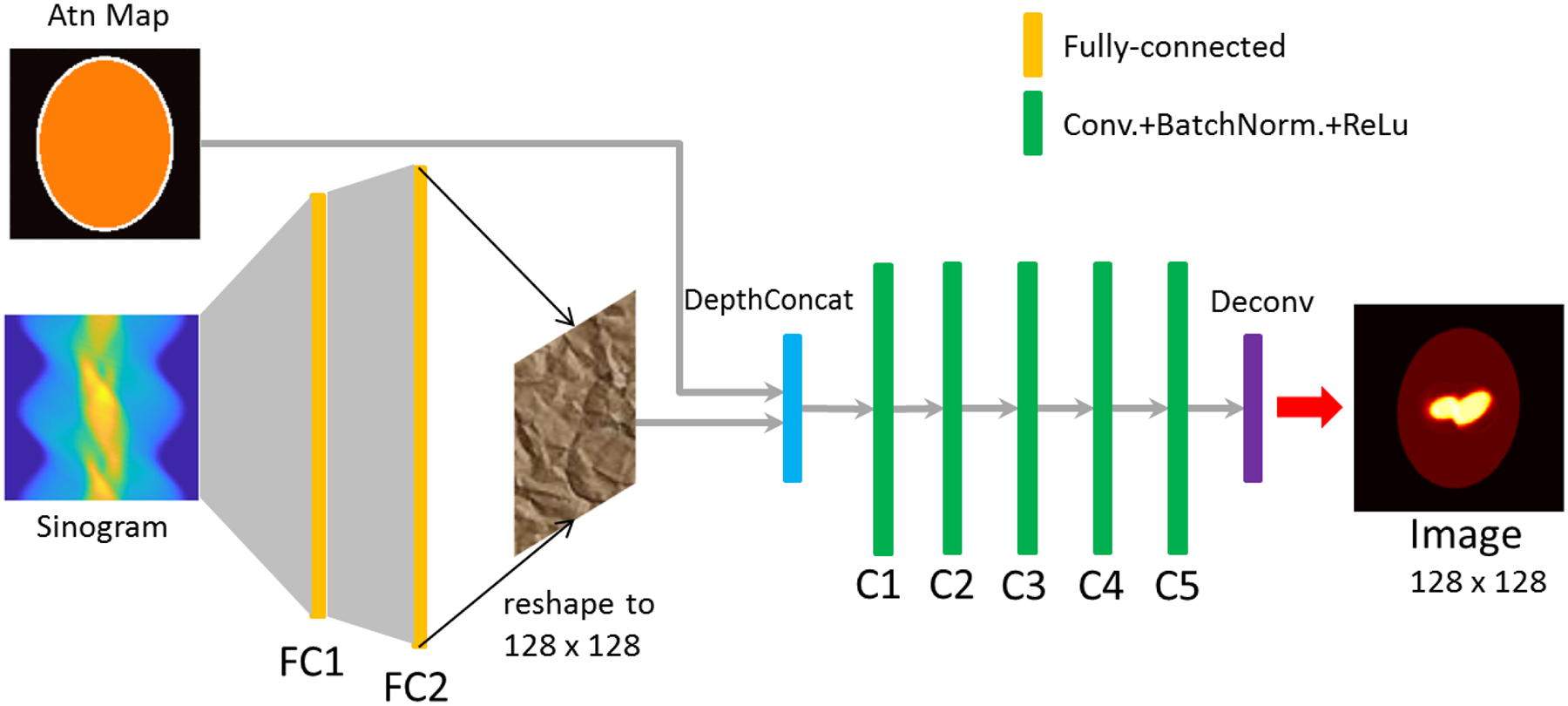
Fig. 1.

The architecture of the proposed deep neural network (DNN) for SPECT medical imaging. The input layer is comprised of two channels to accept the projection data (sinogram) and the attenuation map, respectively. The output of the neural network is the reconstructed activity image. Projection data were fed to the fully connected layers, FC1 and FC2, and the output of FC2 and the attenuation map were together delivered to the subsequent convolutional layers (C1~C5). Each convolutional layer was followed by a batch normalization layer and nonlinear rectifier linear unit (ReLu) function.
