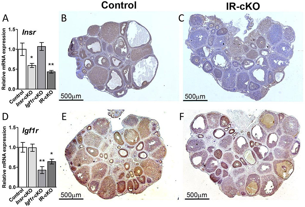
Figure 1.
Conditional ablation of Insr and Igf1r using Pgr-Cre reduces receptor expression in antral follicles and corpora lutea. Samples are from superovulated mice 24 h post-hCG. (A) Insr expression is significantly reduced in total ovarian mRNA from Insr-cKO and IR-cKO mice as assessed by qPCR. Data are expressed as mean ± SEM (n=5, *P <0.05, **P <0.01). (B) Immunohistochemistry (IHC) shows cytoplasmic localization of INSR in granulosa cells of growing follicles and luteal cells of corpora lutea (CL). (C) IHC showing expression of INSR is markedly decreased specifically in the granulosa cell layer of antral follicles and in the CL after conditional ablation of Insr. (D) Igf1r expression is significantly reduced (n=5, *P <0.05, **P <0.01) in total ovarian mRNA from Igf1r-cKO and IR-cKO mice as assessed by qPCR. (E) IHC shows abundant cytoplasmic localization of IGF1R in granulosa cells of growing follicles and in CL. (F) IHC showing expression of IGF1R is markedly decreased specifically in the granulosa cell layer of antral follicles and in the CL after conditional ablation of Insr. IHC images show a representative result obtained in 4 biological repeats.

