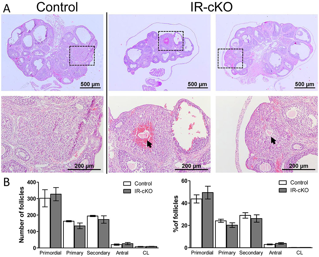
Figure 3.
Histological analysis of IR-cKO ovaries reveals impaired ovulation. Samples are from superovulated mice 24 h post-hCG. (A) Gross analysis of ovarian morphology of IR-cKO mice (2 representative animals) exhibits no decrease in ovarian size. However, all animals exhibit signs of anovulation characterized by oocytes trapped in putative CL tissue (arrows). Boxes indicate the region illustrated by the higher magnification panel below each image. (B) Ovarian follicle distribution is not significantly different in IR-cKO mice compared to controls. Complete ovaries were serially sectioned and numbers of each follicle type scored. Data are presented as mean ± SEM (n=3).

