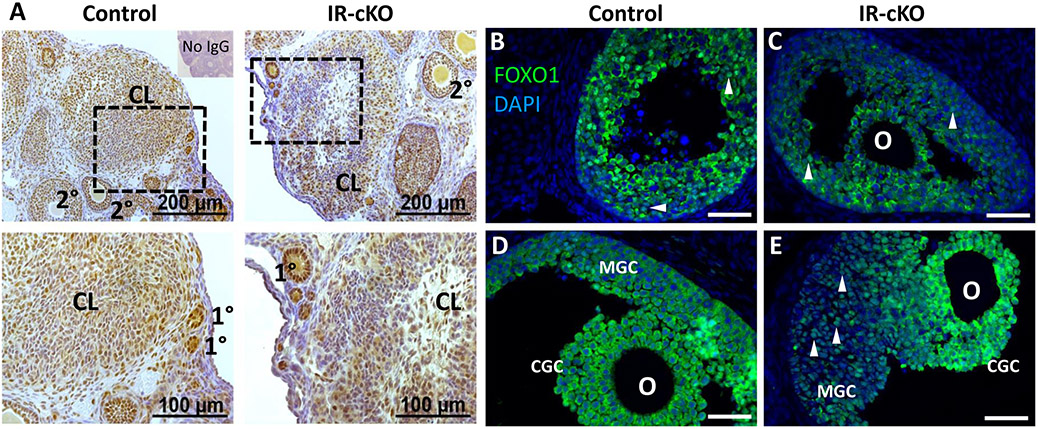
Figure 8.
Insulin receptor signaling mediator AKT1 exhibits reduced phosphorylation in IR-cKO mice. Samples are from superovulated mice 24 h post-hCG. (A) IHC showing diminished phosphorylation of Ser473, a residue of AKT1 activated by insulin receptor stimulation, in a representative animal from each genotype (n=3). Dashed boxes indicate the location of the higher magnification images in the lower panels. Reduced phosphor-AKT1 correlates to large antral follicles and CL where insulin receptors were ablated by Pgr-Cre expression (see Fig. 1) and not globally impaired as primary and secondary follicles retain normal AKT1 activation. (B-E) Immunolocalization of FOXO1, an AKT1-dependent target, is altered in IR-cKO mice. In early antral follicles (B and C), FOXO1 exhibits either nuclear or cytoplasmic localization, present only in granulosa cells, and it does not seem affected by the INSR and IGF1R knockout. (D) In periovulatory follicles of control mice, FOXO1 is translocated to the cytoplasm of both mural granulosa cells (MGC) and cumulus granulosa cells (CGC). (E) However, while the localization of FOXO1 in IR-cKO seems not to be altered in CGC, it is nuclearly localized in MGCs. Arrowheads denote cells with nuclear localization of FOXO1; O-oocyte.

