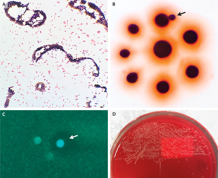
Figure 2. Cerebrospinal Fluid Specimen.
Gram’s staining of the cerebrospinal fluid (Panel A) shows small, round gram-positive organisms. At higher magnification (Panel B), these organisms are of various sizes, well circumscribed, surrounded by a salmon hue, and well separated, with occasional budding on a narrow base (arrow). Calcofluor white staining of a fungal wet preparation (Panel C) shows intense central staining with circumferential clearing (arrow) due to dye exclusion by the polysaccharide capsule. Culture on a blood agar plate (Panel D) shows small, glistening colonies with a mucoid appearance.

