



Abstract
Rationale: Obstructive sleep apnea is recurrent upper airway obstruction caused by a loss of upper airway muscle tone during sleep. The main goal of our study was to determine if designer receptors exclusively activated by designer drugs (DREADD) could be used to activate the genioglossus muscle as a potential novel treatment strategy for sleep apnea. We have previously shown that the prototypical DREADD ligand clozapine-N-oxide increased pharyngeal diameter in mice expressing DREADD in the hypoglossal nucleus. However, the need for direct brainstem viral injections and clozapine-N-oxide toxicity diminished translational potential of this approach, and breathing during sleep was not examined.
Objectives: Here, we took advantage of our model of sleep-disordered breathing in diet-induced obese mice, retrograde properties of the adeno-associated virus serotype 9 (AAV9) viral vector, and the novel DREADD ligand J60.
Methods: We administered AAV9-hSyn-hM3(Gq)-mCherry or control AAV9 into the genioglossus muscle of diet-induced obese mice and examined the effect of J60 on genioglossus activity, pharyngeal patency, and breathing during sleep.
Measurements and Main Results: Compared with control, J60 increased genioglossus tonic activity by greater than sixfold and tongue uptake of 2-deoxy-2-[18F]fluoro-d-glucose by 1.5-fold. J60 increased pharyngeal patency and relieved upper airway obstruction during non-REM sleep.
Conclusions: We conclude that following intralingual administration of AAV9-DREADD, J60 can activate the genioglossus muscle and improve pharyngeal patency and breathing during sleep.
Keywords: chemogenetics, obstructive sleep apnea, pharmacotherapy, hypoglossal motoneurons
At a Glance Commentary
Scientific Knowledge on the Subject
Obstructive sleep apnea (OSA) is recurrent upper airway obstruction caused by a loss of upper airway muscle tone during sleep. OSA is a common disorder affecting 25–30% of the adult population and more than 50% of obese individuals. Continuous positive airway pressure relieves OSA, but poor adherence limits its use. There is no pharmacotherapy for OSA.
What This Study Adds to the Field
The main goal of our study was to determine if designer receptors exclusively activated by designer drugs could be used to activate the genioglossus muscle as a potential novel treatment strategy for sleep apnea.
Obstructive sleep apnea (OSA) is recurrent upper airway obstruction caused by a loss of upper airway muscle tone during sleep (1). OSA is a common disorder affecting 25–30% of the adult population (2, 3) and more than 50% of obese individuals (4, 5). Continuous positive airway pressure relieves OSA, but poor adherence limits its use (6). There is no pharmacotherapy for OSA.
The tongue plays a critical role in maintaining upper airway tone, and it has been targeted for OSA therapy (7–11). The tongue is a unique muscular organ participating in multiple motor functions including speech, chewing, and maintenance of pharyngeal stability. The human tongue contains eight pairs of skeletal muscles. There are four extrinsic muscles, including the genioglossus (GG)—the main pharyngeal dilator and tongue protrudor—and three retractors, the styloglossus, hyoglossus, and palatoglossus, as well as four intrinsic muscles (10). Of these muscles, the GG plays a critical role in defending upper airway patency during sleep (12–15). Motor functions of the tongue are controlled by the hypoglossal nerve originating from the hypoglossal nucleus in the medulla. Multiple potential targets to treat OSA have been identified on hypoglossal motoneurons (HMNs), but efforts to stimulate these motor neurons in humans and animals have had had variable success (16–22).
Designer receptors exclusively activated by designer drugs (DREADD) is a technology that is revolutionizing neuroscience and provides a tool for activating specific neuronal populations. In this regard, we and others have recently demonstrated that viral strategies can be used to drive expression of excitatory DREADD in HMNs. Activation of these DREADD by a ligand clozapine-N-oxide (CNO) increased GG activity as shown by electromyography (EMGGG) (23, 24). Furthermore, dynamic magnetic resonance imaging (MRI) showed that CNO-mediated activation of HMN DREADD improved pharyngeal patency (24). In contrast, when inhibitory DREADD were expressed in HMNs, CNO resulted in inspiratory flow limitation during sleep, a key indicator of upper airway obstruction (25). Although promising as a proof-of-concept, the prior studies of DREADD in HMNs had significant drawbacks that limit the potential clinical application. First, direct brainstem injection of the viral constructs will not be possible in humans. Second, CNO metabolizes to clozapine, an antipsychotic drug with affinity to multiple endogenous receptors (26). Third, brainstem injection of viral constructs is difficult to localize to a discrete region, increasing the possibility of off-target (i.e., non-HMN) injections. Finally, although activation of excitatory DREADD expressed in the hypoglossal nucleus dilated the pharynx and increased GG muscle tone in anesthetized mice, evidence that it treats upper airway obstruction during sleep (i.e., OSA) has not been obtained in any prior study.
The goal of the current study was to develop a novel and minimally invasive gene therapy approach for treating OSA. We used mice with diet-induced obesity (DIO), who exhibit sleep-disordered breathing (SDB) (27). We chose this animal model because our laboratory has demonstrated that mouse upper airway physiology is very similar to humans (28, 29) and that SDB in DIO mice is similar to human disease (27, 30, 31). Prior work has established that the intralingual delivery of adeno-associated virus serotype 9 (AAV9) can effectively drive transgene expression in HMNs (32, 33). Accordingly, we used intralingual delivery of AAV9 encoding excitatory DREADD, hSyn- hM3D (Gq) – mCherry or green fluorescent protein (AAV9-hSyn-GFP) in DIO mice. The injections were targeted to the GG muscle. Four weeks after the infection, we treated mice with a novel and highly specific DREADD ligand, J60 (34), and saline using a crossover study design. Outcome measures included GG EMG activity, tongue metabolic activity, pharyngeal patency by dynamic MRI, and breathing during sleep.
Methods
In total, 27 adult male C57BL/6J mice (Jackson Laboratories) 6–8 weeks of age were fed with a high-fat diet (TD 03584; Teklad, 5.4 kcal/g, 35.2% fat, 58.4% kcal from fat) for 12 weeks and used in our experiments at 18–20 weeks of age. The animals had free access to food and water. Mice were housed in a standard laboratory environment at 22°C in the 12 hours light/dark cycle (09:00 am. to 09:00 pm lights on/09:00 pm. to 09:00 am lights off). All surgical procedures were performed under 1–2% isoflurane anesthesia. Buprenorphine (0.01 mg/kg) was administered subcutaneously at the end of surgery to minimize discomfort. The study was approved by the Johns Hopkins University Animal Use and Care Committee (Protocols # MO16M161 and MO19M161) and complied with the American Physiological Society Guidelines for Animal Studies. The study included four experimental protocols, EMG of the genioglossus muscle of the tongue, dynamic MRI of the upper airway, positron emission tomography scan, and sleep studies. Fourteen mice underwent EMG, MRI, and positron emission tomography scan, and 13 mice underwent EMG followed by sleep studies. Histology of the tongue and the brain was examined in all mice. Detailed description of experimental procedures is provided in the online supplement.
Results
DIO mice treated with DREADD and control virus were of the same age and body weight (Table 1). Both AAV9-hSyn-GFP and AAV9-hSyn-hM3D(Gq)-mCherry were detectable in HMNs (Figures 1A and 1B), which indicates that the viruses administered to the GG muscle traveled retrograde. Moreover, GFP and mCherry were predominately located in the ventral rostral portion of the hypoglossal nucleus (12N), which contains HMNs innervating the GG muscle (35, 36) (Figures 1A and 1B and E1A in the online supplement). In total, 31.7 ± 10% of HMNs were transfected with DREADD. 28.1 ± 9.3% of DREADD-positive HMNs were anatomically located to the retractor area, whereas 71.9 ± 23.9% of DREADD-positive HMNs corresponded to the protrudor compartment. A similar number of HMNs were transfected with GFP control, 33.5 ± 15%, with similar retractor/protrudor distribution, 25.4 ± 3.6% and 74.6 ± 11.3%, respectively. Low levels of GFP and mCherry were present in the GG muscle of the tongue 4–6 weeks after infection (Figures 1C and 1D). No evidence of inflammation was found in the tongue of mice infected with the AAV9-hSyn-GFP and DREADD-containing viruses (Figures 1E and 1F, respectively).
Table 1.
Basic Characteristics of C57BL/6J Mice That Underwent Sleep Studies after Vehicle (Saline) and J60 Treatment
| Treatment | N | Age (wk) | Weight (g) | Temperature (°C) |
|---|---|---|---|---|
| DREADD | 17 | 22.8 ± 1.5 | 45.8 ± 1.5 | 36.2 ± 0.2 |
| GFP | 10 | 23.0 ± 1.6 | 45.3 ± 1.7 | 36.1 ± 0.1 |
Definition of abbreviations: DREADD = designer receptors exclusively activated by designer drugs; GFP = green fluorescent protein.
Values are presented as mean ± SD. No significant difference between groups.
Figure 1.

Histological images in AAV9-hSyn-GFP and AAV-hSyn-DIO-hM3D(Gq)-mCherry designer receptors exclusively activated by designer drugs (DREADD)–treated mice. Fluorescent microscopy images (×20) of the brain medulla show (A) GFP (green fluorescent protein) and (B) DREADD mCherry expression in the hypoglossal nucleus (12 N). Fluorescent images (×20) of the tongue also show (C) GFP and (D) DREADD expression. Cell nuclei were stained with DAPI (blue). Representative hematoxylin and eosin images of the tongue (×20) in (E) GFP- and (F) DREADD-treated mice showed no evidence of inflammation or tissue injury. Scale bars, 50 μm. Arrows represent the localization of DREADD or GFP (n = 27). The mixed effect multivariable linear regression was used for statistical comparisons. 4V = fourth ventricle; Ep = epithelium; M = muscle.
EMGGG measurements in AAV9-hSyn-hM3D(Gq)-mCherry–infected mice showed that J60 increased EMGGG phasic activity by 1.9 ± 0.4-fold (P < 0.001) and induced a striking 6.6 ± 1.6-fold increase (P < 0.001) in EMGGG tonic activity (Figure 2). However, there was no correlation between the percentage of DREADD-transfected HMNs and EMG responses to J60 (Figures E1B–E1D). As expected, saline injections in DREADD-treated mice or J60 injections in mice injected with AAV9-hSyn-GFP virus had no effect on EMGGG. In addition, J60 had no effect on diaphragm EMG in DREADD-treated mice (Figure E2).
Figure 2.

Genioglossus EMG recordings. (A) Representative genioglossal electromyography (EMGGG), moving average (∫EMGGG), and respiratory effort recorded at baseline (left) and after J60 administration (right) in a mouse expressing designer receptors exclusively activated by designer drugs (DREADD) in the hypoglossal motoneurons. Note the robust increase in both phasic and tonic EMG activity after J60. (B) EMG response to J60 or saline in DREADD-treated animals (n = 9; baseline and 15 min after injections) and EMG response to J60 and mice treated with control GFP virus (n = 5, baseline and 15 min after injections). Tonic and phasic EMG values were normalized to peak phasic EMG at baseline and plotted separately. *P < 0.001. The mixed effect multivariable linear regression was used for statistical comparisons. a.u. = arbitrary units; Exp. = expiration; GFP = green fluorescent protein; Insp. = inspiration.
Tongue metabolic activity was assessed using DREADD-assisted metabolic mapping (34). In DREAADD-expressing mice, J60 dramatically increased 2-deoxy-2-[18F]fluoro-d-glucose uptake in the tongue (Figure 3), which indicated an increase in metabolic activity. In contrast, in AAV9-hSyn-GFP–infected mice, J60 had no effect.
Figure 3.

Positron emission tomography data. Representative positron emission tomography scan images (A) in AAV9-hSyn-GFP–treated mice (n = 5) and (B) in AAV-hSyn-DIO-hM3D(Gq)-mCherry–treated mice (n = 7). Note the robust increase in tongue metabolic activity (white arrow) in mice treated with designer receptors exclusively activated by designer drugs (DREADD). 2-Deoxy-2-[18F]fluoro-d-glucose uptake in standardized uptake values in individual mice infected with (C) GFP or (D) DREADD. Bars reflect median values. *P < 0.001. The mixed effect multivariable linear regression was used for statistical comparisons. GFP = green fluorescent protein; SUV = standardized uptake value.
The effect of retrograde AAV9-DREADD on upper airway patency in anesthetized mice was assessed by dynamic MRI as we have previously described (24). J60 significantly increased the cross-sectional area of the velopharynx measured 5 mm caudal to the hard–soft palate junction during inspiration, when respiratory muscles are active. In contrast, more rostral noncollapsible areas were not affected. The J60-induced increase in the pharyngeal area was significantly greater than after saline treatment (P < 0.05, Figure 4).
Figure 4.

Upper airway magnetic resonance imaging was performed after saline and J60 treatments in AAV9-hSyn-hM3D(Gq)-mCherry designer receptors exclusively activated by designer drugs (DREADD)-infected mice (n = 5), whereas only J60 treatment was performed in AAV9-hSyn-GFP–infected mice (n = 4). Representative axial magnetic resonance imaging of the pharynx of the mouse expressing DREADD is shown. The axial sections 5 mm caudal to the hard–soft palate junction after the injection of (A) saline or (B) J60 are shown. Note the dilatation of the upper airway following J60 infusion in comparison to saline (white arrows). (C) Increases in pharynx cross-sectional areas caudal to the hard–soft palate junction on inspiration (Inspiration – Expiration; I-E) before and after treatment were significant only at the 5 mm distance in DREADD-infected mice after J60. *P < 0.05 from baseline, compared with saline in the same mice and compared with J60 in GFP-infected mice. The mixed effect multivariable linear regression was used for statistical comparisons. Scale bar, 1 cm. GFP = green fluorescent protein.
The analysis of sleep studies showed no significant effect of DREADD or J60 on total sleep time, despite shorter time in the chamber in the DREADD group, which inadvertently occurred during our experiments (Table 2). Neither J60 nor vector had an effect on the sleep efficiency, percentage non-REM (NREM) and REM sleep, and number and duration of the sleep bouts.
Table 2.
Sleep Architecture in GFP- and DREADD-infected Mice
| Time in Chamber (min) | Sleep Efficiency (%) | Sleep (min) |
Bouts |
||||||||
|---|---|---|---|---|---|---|---|---|---|---|---|
| Number |
Average Length (min) |
||||||||||
| Total | NREM | % | REM | % | NREM | REM | NREM | REM | |||
| DREADD saline | 331.6 ± 23.5 | 62.8 ± 6.3 | 141.5 ± 14.5 | 132.0 ± 13.9 | 93.3 | 9.4 ± 4.0 | 6.7 | 20 ± 7 | 6 ± 2 | 7.6 ± 4.1 | 1.3 ± 0.5 |
| DREADD J60 | 316.1 ± 34.6 | 66.2 ± 5.2 | 142.5 ± 28.1 | 130.8 ± 23.9 | 91.8 | 11.7 ± 4.9 | 8.2 | 16 ± 4 | 7 ± 3 | 8.6 ± 2.4 | 1.7 ± 0.3 |
| GFP saline | 372.5 ± 17.9 | 72.0 ± 7.3 | 223.4 ± 34.2 | 209.1 ± 34.1 | 93.6 | 14.4 ± 7.6 | 6.4 | 19 ± 3 | 8 ± 4 | 10.8 ± 2.0 | 1.8 ± 0.5 |
| GFP J60 | 386.0 ± 38.3 | 72.2 ± 2.7 | 235.5 ± 34.4 | 221.3 ± 35.8 | 94.0 | 14.1 ± 4.4 | 6.0 | 20 ± 2 | 9 ± 3 | 11.2 ± 2.2 | 1.7 ± 0.2 |
Definition of abbreviations: DREADD = designer receptors exclusively activated by designer drugs; GFP = green fluorescent protein; NREM = non-REM.
Values are presented as mean ± SD unless otherwise noted. Sleep times, number of bouts, and average bout length are shown for GFP- and DREADD-infected mice.
Sleep studies in AAV9-DREADD–infected mice treated with saline showed inspiratory flow limitation, a marker of upper airway obstruction, throughout NREM sleep (Figure 5A), which improved after J60 treatment (Figure 5A). Indeed, maximal inspiratory flow, which is inversely related to the severity of upper airway obstruction (30), increased from 2.1 ± 0.3 ml/min in saline-treated mice to 2.9 ± 0.5 ml/min in J60-treated mice (P < 0.05, Figure 5B). There was also a significant increase in minute ventilation, from 0.59 ± 0.08 to 0.86 ± 0.12 ml/min/kg after saline and J60 treatments, respectively (P < 0.05, Figure 5C). We found no correlation between EMG responses to J60 under anesthesia and increases in maximal inspiratory flow or minute ventilation in response to the DREADD ligand during sleep (not shown). In contrast, the percentage of DREADD-positive HMNs trended to correlate with the J60-induced increase in maximal inspiratory flow (r2 = 0.45, P = 0.06, Figure E1E) and correlated significantly with the J60-induced increase in minute ventilation (r2 = 0.47, P < 0.05, Figure E1F). Although respiratory responses correlated with the total DREADD expression, there was no correlation with DREADD expression in HMNs innervating tongue protrudors or retractors (not shown). AAV9-DREADD–infected mice also showed inspiratory flow limitation during REM sleep (Figure 5F), but J60 did not improve maximal inspiratory flow and e (Figures 5G and 5H). In contrast to the AAV9-DREADD–treated mice, J60 had no effect on inspiratory flow and Ve during sleep in AAV9-hSyn-GFP–treated mice (Figures 5D, 5E, 5I, and 5J).
Figure 5.
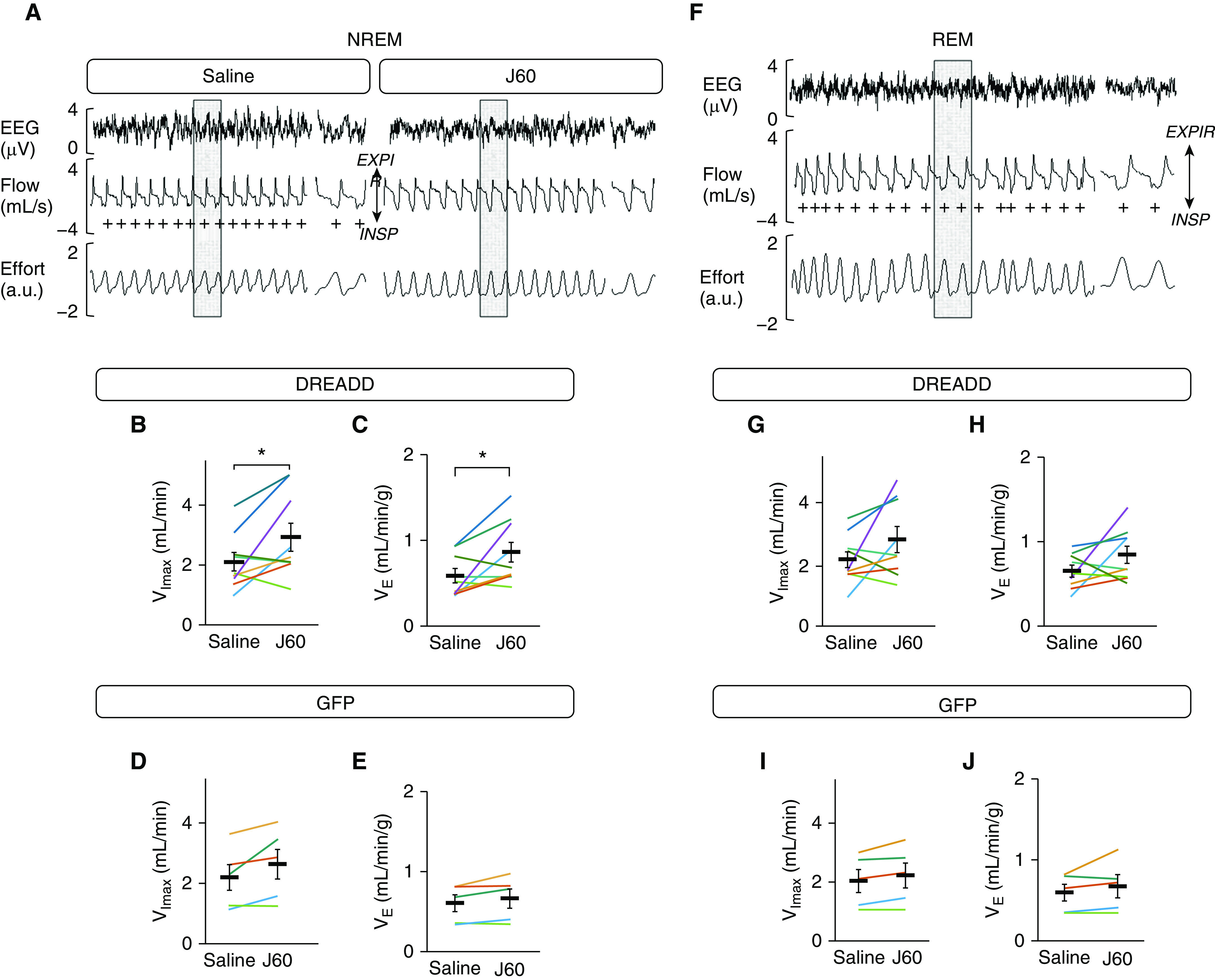
Sleep recordings. (A and F) Representative NREM sleep recordings (A) and REM (F) in the same mouse expressing AAV9-hSyn-hM3D(Gq)-mCherry designer receptors exclusively activated by designer drugs (DREADD) in the hypoglossal motoneurons after treatment with saline (left) versus J60 (right). + denotes breaths with inspiratory flow limitation. For NREM, (B) maximal inspiratory flow (Vimax), (C) e in mice expressing DREADD, and (D) Vimax and (E) e in mice expressing GFP in the hypoglossal motoneurons are shown. For REM, (G) Vimax, (H) e in mice expressing DREADD, and (I) Vimax and (J) e in mice expressing GFP in the hypoglossal motoneurons are shown. Every line in B–J represents an individual mouse treated with saline or J60. Means ± SEM are shown. *P < 0.05 by Wilcoxon rank-sum test. a.u. = arbitrary units; EXPIR = expiration; GFP = green fluorescent protein; INSP = inspiration; NREM = non-REM.
Discussion
The main finding of our study is that intralingual administration of AAV9 encoding excitatory DREADD drives DREADD expression in HMNs and therefore allows control of upper airway patency and improve breathing during sleep in obese mice. The novel and highly potent ligand J60 effectively activated motoneurons containing DREADD and corresponding muscles, which resulted in increased tonic and phasic GG muscle activity and increased inspiratory flow during NREM sleep.
We and others have previously shown that HMNs transfected with AAV-DREADD by stereotactic injections can be remotely controlled with CNO, which modulates GG activity and the pharyngeal diameter (23–25). However, these proof-of-concept studies required brain injections and therefore lacked potential for translation to the pharmacotherapy of OSA. The current study showed that DREADD could be effectively delivered to >30% of HMNs by a retrograde viral vector injected into the GG muscle of the tongue, a major upper airway dilator. Administrations of DREADD with a retrograde vector have been previously used to study brain circuits controlling behavior (37, 38), thermoregulation (39), and axonal regeneration (40). To our knowledge, our study provides the first evidence that DREADD delivery to motoneurons with a retrograde vector administered to a specific muscle can be used to modulate function of this muscle.
Our approach of chemogenetic remote control of the upper airway muscles stands in contrast to previous reports, in which HMNs were manipulated nonselectively. Stereotactic injections of non–Cre-dependent DREADD into the 12N result in DREADD expression specifically in neurons (as opposed to glia) by virtue of the neuron-specific synapsin promoter (41, 42), but this method may result in DREADD expression in any neuron including motoneurons, interneurons, and sensory neurons. Cre-technology enabled investigators to target motoneurons more specifically by administering viral vectors carrying double-floxed inverse open reading frame to Chat-Cre mice (41, 43). In these mice, Cre-recombinase was expressed selectively in cholinergic neurons, such as HMNs (23). However, HMNs are a heterogeneous population innervating tongue protrudors, that is, GG, tongue retractors, and intrinsic lingual muscles. Stimulating the entire population of HMNs can have unpredictable and even counterproductive effects on upper airway patency (44). Similarly, pharmacological stimulation of HMNs based on molecular targets expressed in the entire population of HMNs can also be unpredictable. In contrast, administering the retrograde vector directly into the GG muscle assured >70% of DREADD expression in HMNs innervating the tongue protrudor and pharyngeal dilator muscle, GG, and dilated the pharynx. Although our approach is not as selective as selective electrical stimulation of the hypoglossal nerve (44), it does not require surgical implantation of a pacemaker system.
Our application of chemogenetics is potentially translatable to human OSA and may be relevant for conditions involving neurologic impairment of the pharynx such as stroke, multiple sclerosis, Guillain-Barré syndrome, and others. AAVs are nonpathogenic replication defective vectors, which are safe and efficient instruments for gene delivery (45). AAV-based gene therapy has recently been U.S. Food and Drug Administration–approved for biallelic RPE65 mutation–associated retinal dystrophy (46) and SMN1 mutation–associated spinal muscular atrophy (47), suggesting suitability for other medical therapies as well. Another potentially translatable aspect of our study is a highly potent, inert novel DREADD ligand J60 (34) rather than CNO, which has potential psychotropic activity (26). We provide strong evidence of the J60 high efficiency: J60 induced a greater than sixfold rise in tonic GG EMG activity and a dramatic increase in tongue muscle metabolic activity in DREADD-infected mice.
A major advantage and unique aspect of our approach is that we demonstrated improvements in multiple outcomes including pharyngeal patency, inspiratory flow, and e during sleep with DREADD activation. Rather than limiting our outcomes to the tongue muscle activity, responses in these additional outcomes reinforce the concept that the J60 ligand produced clinically relevant improvements of pharyngeal stability. Indeed, clinical studies of the hypoglossal nerve stimulation in OSA have previously demonstrated that an increase in tongue muscle activity may lead to pharyngeal closure and apneas if the “wrong” muscles (i.e., tongue retractors only) are activated (44). Here, we have shown that selective stimulation of HMNs with J60 dilated the most collapsible segment of the pharynx (48), whereas noncollapsible segments adjacent to the hard palate were not affected, and that the pharyngeal dilatation increased maximal inspiratory flow and e in NREM sleep. Notably, neither DREADD nor J60 impaired the quality of sleep as evident from similar numbers and durations of NREM and REM sleep bouts.
Our data showed no correlation between the percentage of DREADD-positive HMNs and the tongue muscle activity in response to J60 and between EMG and respiratory responses to J60. We attribute this finding to the fact that EMG was measured under isoflurane anesthesia, which may have a variable effect on tongue muscle phasic and tonic activity (49). In contrast, total DREADD expression in HMNs positively correlated with respiratory augmentation by J60 during NREM sleep. In addition, although the percentage of all DREADD-positive HMNs correlated with breathing parameters during sleep, there was no relationship between DREADD expression in protrudors or retractors and breathing responses to J60. The latter can be related either to an investigator error in identification of HMN groups or to an insufficient number of observations. Alternatively, this finding may be of physiological importance. It is conceivable that combined stimulation of protrudors and retractors is necessary to open upper airway during sleep, as suggested by previous animal and human research (44, 50–52).
Our study provides definitive evidence that chemogenetic stimulation of HMNs innervating GG relieves airway obstruction and hypoventilation in mice with SDB. The lack of effect of J60 on the diaphragm further confirms that our intervention was specific for upper airway patency. J60 did not improve pharyngeal patency in REM sleep, which is characterized by profound inhibition of GG activity (53). Horton and colleagues (23) reported that chemogenetic manipulation increased tongue muscle activity during REM sleep, but DREADD was delivered directly to the hypoglossal nucleus by brain injection. It is conceivable that in our study retrograde administration did not deliver a sufficient amount of the Gq receptor to GG motoneurons to elicit protective responses in REM sleep. Alternatively, simultaneous to GG recruitment, other pharyngeal muscles, that is, tongue retractors, intrinsic tongue muscles, and nonlingual muscles, which was not achieved by our approach, may be particularly important in REM sleep (54, 55).
The study has several limitations. First, SDB in DIO mice is more consistent with obesity hypoventilation with continuous inspiratory flow limitation due to pharyngeal obstruction rather than typical OSA with distinct obstructive apneas (27). However, this pattern of SDB was described in obese premenopausal women (56) and in children (57, 58), and therefore, DIO mice can serve as an animal model for therapeutic development in OSA. Second, although muscle injection of the DREADD replaced brain injections, the virus must be deployed in the brain to be effective. As we described above, AAVs are safe and self-limited (46). AAV is not integrated in the genome and its lifespan in the neuron is short, which greatly diminishes a risk of serious adverse outcomes (59). Nevertheless, human use of AAV9-DREADD may be limited to very severe life-threatening OSA in patients who are noncompliant with continuous positive airway pressure or in patients with severe neurological impairment. Third, AAV9 is a relatively inefficient retrograde vector (60) (Figure 1). However, even low levels of AAV9 infection had a dramatic effect on upper airway patency and SDB in NREM sleep. AAV9 also possess limited anterograde properties (60), which may result in undesirable spread to sensory gustatory or tactile neurons. Alternative AAV vectors with stronger and more specific retrograde properties should be tested in the future. Fourth, J60 had no effect on SDB during REM sleep, which could be related to the low AAV9-DREADD transfection rate or the lack of activation of other than GG muscles.
In conclusion, our study provided initial evidence that administration of retrograde AAV9-DREADD to the GG muscle restores pharyngeal patency and treats SDB, which may open new horizons for OSA therapy.
Supplementary Material
Footnotes
Supported by NIH grants R01HL128970 and R01HL138932 to V.Y.P. and R01HL144859 to A.R.S. and by the American Heart Association grant AHA 19CDA34660245 and an American Thoracic Society unrestricted award to T.F.C.
Author Contributions: T.F.C., L.U.S., A.R.S., D.D.F., and V.Y.P. conceptualized the work. T.F.C., K.F., R.G.S., M.M., and V.Y.P. designed the experiments. T.F.C., H.P., C.F., M.R.A., J.B., L.J.K., R.L., M.E.C., K.F., and M.J.B. performed the experiments. T.F.C., H.P., C.F., J.B., R.L., S.R.S., K.F., M.J.B., M.M., and V.Y.P. analyzed the data. T.F.C., H.P., and M.J.B. did the statistical analysis. J.B., M.M., and D.D.F. provided reagents. T.F.C., M.R.A., J.B., L.G.B., L.U.S., K.F., R.G.S., A.R.S., M.J.B., M.M., D.D.F., and V.Y.P. wrote the manuscript.
This article has an online supplement, which is accessible from this issue’s table of contents at www.atsjournals.org.
Originally Published in Press as DOI: 10.1164/rccm.202002-0321OC on July 16, 2020
Author disclosures are available with the text of this article at www.atsjournals.org.
References
- 1.Gastaut H, Tassinari CA, Duron B. Polygraphic study of diurnal and nocturnal (hypnic and respiratory) episodal manifestations of Pickwick syndrome [in French] Rev Neurol (Paris) 1965;112:568–579. [PubMed] [Google Scholar]
- 2.Peppard PE, Young T, Barnet JH, Palta M, Hagen EW, Hla KM. Increased prevalence of sleep-disordered breathing in adults. Am J Epidemiol. 2013;177:1006–1014. doi: 10.1093/aje/kws342. [DOI] [PMC free article] [PubMed] [Google Scholar]
- 3.Heinzer R, Vat S, Marques-Vidal P, Marti-Soler H, Andries D, Tobback N, et al. Prevalence of sleep-disordered breathing in the general population: the HypnoLaus study. Lancet Respir Med. 2015;3:310–318. doi: 10.1016/S2213-2600(15)00043-0. [DOI] [PMC free article] [PubMed] [Google Scholar]
- 4.Tufik S, Santos-Silva R, Taddei JA, Bittencourt LR. Obstructive sleep apnea syndrome in the Sao Paulo epidemiologic sleep study. Sleep Med. 2010;11:441–446. doi: 10.1016/j.sleep.2009.10.005. [DOI] [PubMed] [Google Scholar]
- 5.Punjabi NM, Bandeen-Roche K, Marx JJ, Neubauer DN, Smith PL, Schwartz AR. The association between daytime sleepiness and sleep-disordered breathing in NREM and REM sleep. Sleep. 2002;25:307–314. [PubMed] [Google Scholar]
- 6.Kribbs NB, Pack AI, Kline LR, Smith PL, Schwartz AR, Schubert NM, et al. Objective measurement of patterns of nasal CPAP use by patients with obstructive sleep apnea. Am Rev Respir Dis. 1993;147:887–895. doi: 10.1164/ajrccm/147.4.887. [DOI] [PubMed] [Google Scholar]
- 7.White DP. Pharmacologic approaches to the treatment of obstructive sleep apnea. Sleep Med Clin. 2016;11:203–212. doi: 10.1016/j.jsmc.2016.01.007. [DOI] [PubMed] [Google Scholar]
- 8.Schwartz AR, Smith PL, Oliven A. Electrical stimulation of the hypoglossal nerve: a potential therapy. J Appl Physiol (1985) 2014;116:337–344. doi: 10.1152/japplphysiol.00423.2013. [DOI] [PMC free article] [PubMed] [Google Scholar]
- 9.Strollo PJ, Jr, Soose RJ, Maurer JT, de Vries N, Cornelius J, Froymovich O, et al. STAR Trial Group. Upper-airway stimulation for obstructive sleep apnea. N Engl J Med. 2014;370:139–149. doi: 10.1056/NEJMoa1308659. [DOI] [PubMed] [Google Scholar]
- 10.Zaidi FN, Meadows P, Jacobowitz O, Davidson TM. Tongue anatomy and physiology, the scientific basis for a novel targeted neurostimulation system designed for the treatment of obstructive sleep apnea. Neuromodulation. 2013;16:376–386. doi: 10.1111/j.1525-1403.2012.00514.x. [Discussion, p. 386.] [DOI] [PubMed] [Google Scholar]
- 11.Grace KP, Hughes SW, Horner RL. Identification of the mechanism mediating genioglossus muscle suppression in REM sleep. Am J Respir Crit Care Med. 2013;187:311–319. doi: 10.1164/rccm.201209-1654OC. [DOI] [PubMed] [Google Scholar]
- 12.Malhotra A, Pillar G, Fogel RB, Edwards JK, Ayas N, Akahoshi T, et al. Pharyngeal pressure and flow effects on genioglossus activation in normal subjects. Am J Respir Crit Care Med. 2002;165:71–77. doi: 10.1164/ajrccm.165.1.2011065. [DOI] [PubMed] [Google Scholar]
- 13.Eckert DJ, McEvoy RD, George KE, Thomson KJ, Catcheside PG. Genioglossus reflex inhibition to upper-airway negative-pressure stimuli during wakefulness and sleep in healthy males. J Physiol. 2007;581:1193–1205. doi: 10.1113/jphysiol.2007.132332. [DOI] [PMC free article] [PubMed] [Google Scholar]
- 14.Younes M, Loewen AH, Ostrowski M, Laprairie J, Maturino F, Hanly PJ. Genioglossus activity available via non-arousal mechanisms vs. that required for opening the airway in obstructive apnea patients. J Appl Physiol (1985) 2012;112:249–258. doi: 10.1152/japplphysiol.00312.2011. [DOI] [PubMed] [Google Scholar]
- 15.Dempsey JA, Veasey SC, Morgan BJ, O’Donnell CP. Pathophysiology of sleep apnea. Physiol Rev. 2010;90:47–112. doi: 10.1152/physrev.00043.2008. [DOI] [PMC free article] [PubMed] [Google Scholar]
- 16.Torontali ZA, Grace KP, Horner RL, Peever JH. Cholinergic involvement in control of REM sleep paralysis. J Physiol. 2014;592:1425–1426. doi: 10.1113/jphysiol.2014.271304. [DOI] [PMC free article] [PubMed] [Google Scholar]
- 17.Grace KP, Hughes SW, Horner RL. Identification of a pharmacological target for genioglossus reactivation throughout sleep. Sleep (Basel) 2014;37:41–50. doi: 10.5665/sleep.3304. [DOI] [PMC free article] [PubMed] [Google Scholar]
- 18.Carley DW, Olopade C, Ruigt GS, Radulovacki M. Efficacy of mirtazapine in obstructive sleep apnea syndrome. Sleep. 2007;30:35–41. doi: 10.1093/sleep/30.1.35. [DOI] [PubMed] [Google Scholar]
- 19.Song G, Poon C-S. α2-Adrenergic blockade rescues hypoglossal motor defense against obstructive sleep apnea. JCI Insight. 2017;2:e91456. doi: 10.1172/jci.insight.91456. [DOI] [PMC free article] [PubMed] [Google Scholar]
- 20.Taranto-Montemurro L, Edwards BA, Sands SA, Marques M, Eckert DJ, White DP, et al. Desipramine increases genioglossus activity and reduces upper airway collapsibility during non-REM sleep in healthy subjects. Am J Respir Crit Care Med. 2016;194:878–885. doi: 10.1164/rccm.201511-2172OC. [DOI] [PMC free article] [PubMed] [Google Scholar]
- 21.Taranto-Montemurro L, Messineo L, Sands SA, Azarbarzin A, Marques M, Edwards BA, et al. The combination of atomoxetine and oxybutynin greatly reduces obstructive sleep apnea severity: a randomized, placebo-controlled, double-blind crossover trial. Am J Respir Crit Care Med. 2019;199:1267–1276. doi: 10.1164/rccm.201808-1493OC. [DOI] [PMC free article] [PubMed] [Google Scholar]
- 22.Fleury Curado T, Berger S, Polotsky VY. Pharmacotherapy of obstructive sleep apnea: is salvation just around a corner? Am J Respir Crit Care Med. 2019;199:1186–1187. doi: 10.1164/rccm.201811-2135ED. [DOI] [PMC free article] [PubMed] [Google Scholar]
- 23.Horton GA, Fraigne JJ, Torontali ZA, Snow MB, Lapierre JL, Liu H, et al. Activation of the hypoglossal to tongue musculature motor pathway by remote control. Sci Rep. 2017;7:45860. doi: 10.1038/srep45860. [DOI] [PMC free article] [PubMed] [Google Scholar]
- 24.Fleury Curado T, Fishbein K, Pho H, Brennick M, Dergacheva O, Sennes LU, et al. Chemogenetic stimulation of the hypoglossal neurons improves upper airway patency. Sci Rep. 2017;7:44392. doi: 10.1038/srep44392. [DOI] [PMC free article] [PubMed] [Google Scholar]
- 25.Fleury Curado TA, Pho H, Dergacheva O, Berger S, Lee R, Freire C, et al. Silencing of hypoglossal motoneurons leads to sleep disordered breathing in lean mice. Front Neurol. 2018;9:962. doi: 10.3389/fneur.2018.00962. [DOI] [PMC free article] [PubMed] [Google Scholar]
- 26.Gomez JL, Bonaventura J, Lesniak W, Mathews WB, Sysa-Shah P, Rodriguez LA, et al. Chemogenetics revealed: DREADD occupancy and activation via converted clozapine. Science. 2017;357:503–507. doi: 10.1126/science.aan2475. [DOI] [PMC free article] [PubMed] [Google Scholar]
- 27.Fleury Curado T, Pho H, Berger S, Caballero-Eraso C, Shin M-K, Sennes LU, et al. Sleep-disordered breathing in C57BL/6J mice with diet-induced obesity. Sleep (Basel) 2018;41:zsy089. doi: 10.1093/sleep/zsy089. [DOI] [PMC free article] [PubMed] [Google Scholar]
- 28.Polotsky M, Elsayed-Ahmed AS, Pichard L, Richardson RA, Smith PL, Schneider H, et al. Effect of age and weight on upper airway function in a mouse model. J Appl Physiol (1985) 2011;111:696–703. doi: 10.1152/japplphysiol.00123.2011. [DOI] [PMC free article] [PubMed] [Google Scholar]
- 29.Polotsky M, Elsayed-Ahmed AS, Pichard L, Harris CC, Smith PL, Schneider H, et al. Effects of leptin and obesity on the upper airway function. J Appl Physiol (1985) 2012;112:1637–1643. doi: 10.1152/japplphysiol.01222.2011. [DOI] [PMC free article] [PubMed] [Google Scholar]
- 30.Pho H, Hernandez AB, Arias RS, Leitner EB, Van Kooten S, Kirkness JP, et al. The effect of leptin replacement on sleep-disordered breathing in the leptin-deficient ob/ob mouse. J Appl Physiol (1985) 2016;120:78–86. doi: 10.1152/japplphysiol.00494.2015. [DOI] [PMC free article] [PubMed] [Google Scholar]
- 31.Yao Q, Pho H, Kirkness J, Ladenheim EE, Bi S, Moran TH, et al. Localizing effects of leptin on upper airway and respiratory control during sleep. Sleep (Basel) 2016;39:1097–1106. doi: 10.5665/sleep.5762. [DOI] [PMC free article] [PubMed] [Google Scholar]
- 32.Doyle BM, Turner SMF, Sunshine MD, Doerfler PA, Poirier AE, Vaught LA, et al. AAV gene therapy utilizing glycosylation-independent lysosomal targeting tagged GAA in the hypoglossal motor system of pompe mice. Mol Ther Methods Clin Dev. 2019;15:194–203. doi: 10.1016/j.omtm.2019.08.009. [DOI] [PMC free article] [PubMed] [Google Scholar]
- 33.Elmallah MK, Falk DJ, Nayak S, Federico RA, Sandhu MS, Poirier A, et al. Sustained correction of motoneuron histopathology following intramuscular delivery of AAV in pompe mice. Mol Ther. 2014;22:702–712. doi: 10.1038/mt.2013.282. [DOI] [PMC free article] [PubMed] [Google Scholar]
- 34.Bonaventura J, Eldridge MAG, Hu F, Gomez JL, Sanchez-Soto M, Abramyan AM, et al. High-potency ligands for DREADD imaging and activation in rodents and monkeys. Nat Commun. 2019;10:4627. doi: 10.1038/s41467-019-12236-z. [DOI] [PMC free article] [PubMed] [Google Scholar]
- 35.Dobbins EG, Feldman JL. Differential innervation of protruder and retractor muscles of the tongue in rat. J Comp Neurol. 1995;357:376–394. doi: 10.1002/cne.903570305. [DOI] [PubMed] [Google Scholar]
- 36.McClung JR, Goldberg SJ. Organization of the hypoglossal motoneurons that innervate the horizontal and oblique components of the genioglossus muscle in the rat. Brain Res. 2002;950:321–324. doi: 10.1016/s0006-8993(02)03240-7. [DOI] [PubMed] [Google Scholar]
- 37.Li H, Illenberger JM, Cranston MN, Mactutus CF, McLaurin KA, Harrod SB, et al. Posterior ventral tegmental area-nucleus accumbens shell circuitry modulates response to novelty. PLoS One. 2019;14:e0213088. doi: 10.1371/journal.pone.0213088. [DOI] [PMC free article] [PubMed] [Google Scholar]
- 38.Augur IF, Wyckoff AR, Aston-Jones G, Kalivas PW, Peters J. Chemogenetic activation of an extinction neural circuit reduces cue-induced reinstatement of cocaine seeking. J Neurosci. 2016;36:10174–10180. doi: 10.1523/JNEUROSCI.0773-16.2016. [DOI] [PMC free article] [PubMed] [Google Scholar]
- 39.de Git KCG, van Tuijl DC, Luijendijk MCM, Wolterink-Donselaar IG, Ghanem A, Conzelmann K-K, et al. Anatomical projections of the dorsomedial hypothalamus to the periaqueductal grey and their role in thermoregulation: a cautionary note. Physiol Rep. 2018;6:e13807. doi: 10.14814/phy2.13807. [DOI] [PMC free article] [PubMed] [Google Scholar]
- 40.Jaiswal PB, Mistretta OC, Ward PJ, English AW. Chemogenetic enhancement of axon regeneration following peripheral nerve injury in the SLICK-A mouse. Brain Sci. 2018;8:93. doi: 10.3390/brainsci8050093. [DOI] [PMC free article] [PubMed] [Google Scholar]
- 41.Roth BL. DREADDs for neuroscientists. Neuron. 2016;89:683–694. doi: 10.1016/j.neuron.2016.01.040. [DOI] [PMC free article] [PubMed] [Google Scholar]
- 42.Zhu H, Roth BL. DREADD: a chemogenetic GPCR signaling platform. Int J Neuropsychopharmacol. 2014;18:pyu007. doi: 10.1093/ijnp/pyu007. [DOI] [PMC free article] [PubMed] [Google Scholar]
- 43.Zhu H, Aryal DK, Olsen RH, Urban DJ, Swearingen A, Forbes S, et al. Cre-dependent DREADD (designer receptors exclusively activated by designer drugs) mice. Genesis. 2016;54:439–446. doi: 10.1002/dvg.22949. [DOI] [PMC free article] [PubMed] [Google Scholar]
- 44.Fleury Curado T, Oliven A, Sennes LU, Polotsky VY, Eisele D, Schwartz AR. Neurostimulation treatment of OSA. Chest. 2018;154:1435–1447. doi: 10.1016/j.chest.2018.08.1070. [DOI] [PMC free article] [PubMed] [Google Scholar]
- 45.Pattali R, Mou Y, Li X-J. AAV9 vector: a novel modality in gene therapy for spinal muscular atrophy. Gene Ther. 2019;26:287–295. doi: 10.1038/s41434-019-0085-4. [DOI] [PubMed] [Google Scholar]
- 46.Rodrigues GA, Shalaev E, Karami TK, Cunningham J, Slater NKH, Rivers HM. Pharmaceutical development of AAV-based gene therapy products for the eye. Pharm Res. 2018;36:29. doi: 10.1007/s11095-018-2554-7. [DOI] [PMC free article] [PubMed] [Google Scholar]
- 47.Mahajan R. Onasemnogene abeparvovec for spinal muscular atrophy: the costlier drug ever. Int J Appl Basic Med Res. 2019;9:127–128. doi: 10.4103/ijabmr.IJABMR_190_19. [DOI] [PMC free article] [PubMed] [Google Scholar]
- 48.Brennick MJ, Pickup S, Dougherty L, Cater JR, Kuna ST. Pharyngeal airway wall mechanics using tagged magnetic resonance imaging during medial hypoglossal nerve stimulation in rats. J Physiol. 2004;561:597–610. doi: 10.1113/jphysiol.2004.073502. [DOI] [PMC free article] [PubMed] [Google Scholar]
- 49.Eikermann M, Malhotra A, Fassbender P, Zaremba S, Jordan AS, Gautam S, et al. Differential effects of isoflurane and propofol on upper airway dilator muscle activity and breathing. Anesthesiology. 2008;108:897–906. doi: 10.1097/ALN.0b013e31816c8a60. [DOI] [PMC free article] [PubMed] [Google Scholar]
- 50.Fuller D, Mateika JH, Fregosi RF. Co-activation of tongue protrudor and retractor muscles during chemoreceptor stimulation in the rat. J Physiol. 1998;507:265–276. doi: 10.1111/j.1469-7793.1998.265bu.x. [DOI] [PMC free article] [PubMed] [Google Scholar]
- 51.Fuller D, Williams JS, Janssen PL, Fregosi RF. Effect of co-activation of tongue protrudor and retractor muscles on tongue movements and pharyngeal airflow mechanics in the rat. J Physiol. 1999;519:601–613. doi: 10.1111/j.1469-7793.1999.0601m.x. [DOI] [PMC free article] [PubMed] [Google Scholar]
- 52.Oliven A, Odeh M, Geitini L, Oliven R, Steinfeld U, Schwartz AR, et al. Effect of co-activation of tongue protrusor and retractor muscles on pharyngeal lumen and airflow in sleep apnea patients. J Appl Physiol. 1985;2007:1662–1668. doi: 10.1152/japplphysiol.00620.2007. [DOI] [PubMed] [Google Scholar]
- 53.Aggarwal JA, Liu W-Y, Montandon G, Liu H, Hughes SW, Horner RL. Measurement and state-dependent modulation of hypoglossal motor excitability and responsivity in-vivo. Sci Rep. 2020;10:550. doi: 10.1038/s41598-019-57328-4. [DOI] [PMC free article] [PubMed] [Google Scholar]
- 54.Dotan Y, Pillar G, Schwartz AR, Oliven A. Asynchrony of lingual muscle recruitment during sleep in obstructive sleep apnea. J Appl Physiol (1985) 2015;118:1516–1524. doi: 10.1152/japplphysiol.00937.2014. [DOI] [PubMed] [Google Scholar]
- 55.Oliven R, Cohen G, Dotan Y, Somri M, Schwartz AR, Oliven A. Alteration in upper airway dilator muscle coactivation during sleep: comparison of patients with obstructive sleep apnea and healthy subjects. J Appl Physiol (1985) 2018;124:421–429. doi: 10.1152/japplphysiol.01067.2016. [DOI] [PMC free article] [PubMed] [Google Scholar]
- 56.Bonsignore MR, Saaresranta T, Riha RL. Sex differences in obstructive sleep apnoea. Eur Respir Rev. 2019;28:190030. doi: 10.1183/16000617.0030-2019. [DOI] [PMC free article] [PubMed] [Google Scholar]
- 57.Marcus CL. Sleep-disordered breathing in children. Am J Respir Crit Care Med. 2001;164:16–30. doi: 10.1164/ajrccm.164.1.2008171. [DOI] [PubMed] [Google Scholar]
- 58.Marcus CL, McColley SA, Carroll JL, Loughlin GM, Smith PL, Schwartz AR. Upper airway collapsibility in children with obstructive sleep apnea syndrome. J Appl Physiol (1985) 1994;77:918–924. doi: 10.1152/jappl.1994.77.2.918. [DOI] [PubMed] [Google Scholar]
- 59.Naso MF, Tomkowicz B, Perry WL, III, Strohl WR. Adeno-associated virus (AAV) as a vector for gene therapy. BioDrugs. 2017;31:317–334. doi: 10.1007/s40259-017-0234-5. [DOI] [PMC free article] [PubMed] [Google Scholar]
- 60.Castle MJ, Gershenson ZT, Giles AR, Holzbaur ELF, Wolfe JH. Adeno-associated virus serotypes 1, 8, and 9 share conserved mechanisms for anterograde and retrograde axonal transport. Hum Gene Ther. 2014;25:705–720. doi: 10.1089/hum.2013.189. [DOI] [PMC free article] [PubMed] [Google Scholar]
Associated Data
This section collects any data citations, data availability statements, or supplementary materials included in this article.