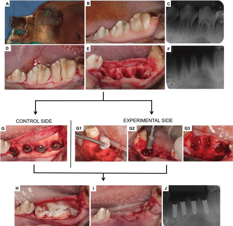
Figure 1.
Surgical procedures sequence: A) Animal under general anesthesia; B) Initial clinical aspect of the premolar teeth; C) Initial radiographic features; D) Sagittal section; E) Fresh alveolus; F) Post-extraction radiographic features; G) Immediately-placed implants on the control side; G1) BM-MSCs+PRP gel; G2-G3) Implants + BM-MSCs+PRP on the experimental side; H) Placement of the collagen membrane; F) Suture and J) Final radiographic features.

