
Figure 4. Comparison of CAR designs.
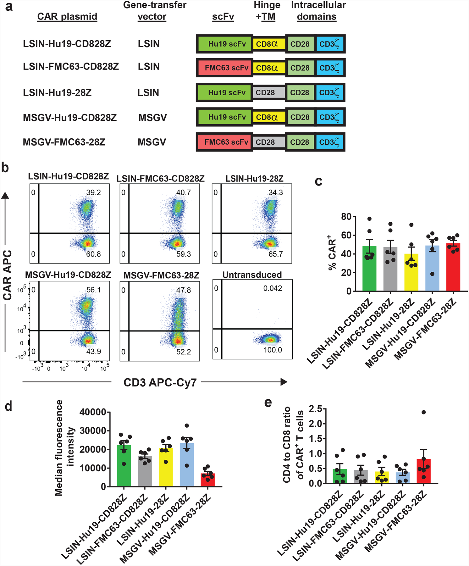
(a) Five CAR plasmids are listed. LSIN, lentiviral vector; MSGV, gamma-retroviral vector. All 5 CARs contained a CD28 costimulatory domain and a CD3ζ domain. LSIN-Hu19-CD828Z had the Hu19 human scFv plus CD8α hinge and transmembrane (TM) domains. LSIN-FMC63-CD828Z had the FMC63 scFv plus CD8α hinge and transmembrane domains. LSIN-Hu19–28Z had the Hu19 scFv plus CD28 hinge and transmembrane domains. MSGV-Hu19-CD828Z had the Hu19 scFv plus CD8α hinge and transmembrane domains; MSGV-Hu19-CD828Z was identical to LSIN-Hu19-CD828Z except for the gene-therapy vectors. MSGV-FMC63–28Z had the murine FMC63 scFv plus CD28 hinge and transmembrane domains. (b) T cells from the same patient were transduced with each of 5 CARs or left untransduced as indicated. Plots are gated on live, CD3+ lymphocytes. These results are representative of results from 6 unique donors. (c) The %CAR+ T cells for each of the 5 CARs is shown. Flow cytometry gating was as in b. LSIN-Hu19-CD828Z was compared to the other 4 CARs. The only consistent difference was between LSIN-Hu19-CD828Z and LSIN-Hu19–28Z (P=0.031). LSIN-Hu19-CD828Z was compared to the other 3 CARs; P values for the comparisons were: LSIN-FMC63-CD828Z, 0.6875; MSGV-Hu19-CD828Z, >0.999; MSGV-FMC63–28Z, 0.5625. (d) The median fluorescence intensities of only the CAR+ T cells are shown. When LSIN-Hu19-CD828Z was compared to the other 4 CARs, the only consistent difference was between LSIN-Hu19-CD828Z and MSGV-FMC63–28Z (P=0.031). LSIN-Hu19-CD828Z was compared to the other 3 CARs; P values for the comparisons were: LSIN-FMC63-CD828Z, 0.063; LSIN-Hu19–28Z, 0.4375; MSGV-Hu19-CD828Z, >0.999. (e) A CD4+ versus CD8+ plot gated on CD3+CAR+ events was used to determine the CD4 to CD8 ratio of CAR+ T cells. LSIN-Hu19-CD828Z was compared to the other 4 CARs; P values for the comparisons were: LSIN-FMC63-CD828Z, 0.438; LSIN-Hu19–28Z, 0.438; MSGV-Hu19-CD828Z, 0.563; MSGV-FMC63–28Z, 0.094. For c-e, comparisons were by 2-tailed Wilcoxon matched-pairs signed-rank test. Graphs c-e show mean +/−SEM. For c-e, n=6 independent experiments with cells from unique donors.