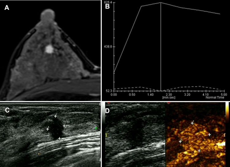
Figure 2.
A 35-year-old female patient found a breast lump in the right outer lower quadrant when she made a breast screening. The examination was normal. (A) DCE-MRI reported mass-like hyperenhancement. (B) The TIC curve showed wash-out type. (C) The US presented irregular hypoechoic lesion sized 6*8 mm, and the surrounding tissues were tangled. (D) CEUS showed homogeneous enhancement and hyperenhancement. After the enhancement, the border slightly expanded. The pathology result is invasive ductal carcinoma (IDC).

