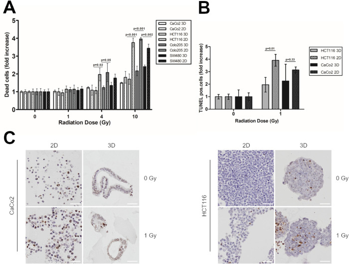
Fig 3. 3D spheroids show decreased apoptosis rates after irradiation compared with 2D cultures.
(A) CRC cells were irradiated with 1, 4, and 10 Gy. Six days after irradiation, cells were stained with annexin V/ PI, and analysed by flow cytometry. Significance was calculated using a two-sided, unpaired Student’s T test. (B) CaCo2 and HCT116 cells were irradiated with 0 and 1 Gy. Six days after irradiation, cells were fixed with 4% PFA, embedded in paraffin and stained for TUNEL assay. Significance was calculated using a two-sided, unpaired Student’s T test. (C) Representative TUNEL stainings for CaCo2 and HCT116 2D and 3D cultures six days after irradiation with 0 and 1 Gy. Scale bar 50 μm.

