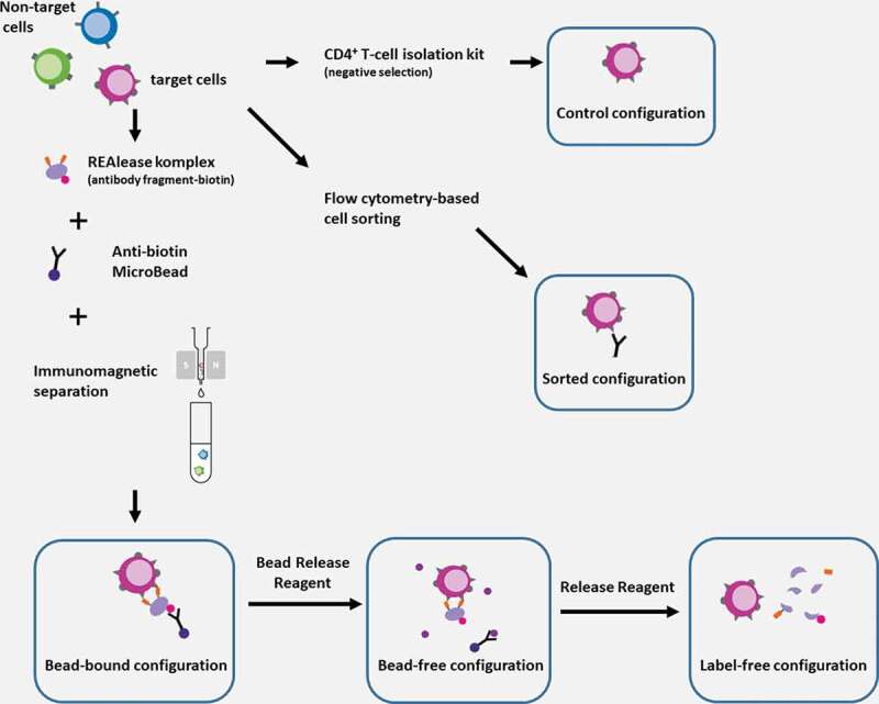
Figure 1.

Schematic illustration of cell separation techniques and the resulting configurations
In vitro activated PBMC samples contain CD4+ “target” cells (purple) to be separated from the other, “non-target” cells (blue and green) in the cell mixture. Separation was accomplished using three different techniques resulting five experimental configurations. In the first case, separation was made with REAlease® CD4 MicroBead Kit resulting the bead-bound configuration (bottom-left). After removing the anti-biotin antibody-microbead conjugate from the cells using Bead Release Reagent, the bead-free configuration was achieved (bottom-middle). By removing the REAlease complex form the separated cell’s surface with Release Reagent, we realized the label free configuration (bottom-right). As control, we used a CD4+ T cell isolation kit, that provides a negative selection and thus, isolated cell were not labeled by any antibody (control configuration, top right). A conventional flow cytometry-based cell sorting using positive selection was applied to achieve the sorted configuration (middle-right). Separated cells were subsequently used for experiments. Some parts of the figure were adapted from the original product descriptions of the REAlease® CD4 MicroBead Kit (with the permission of Miltenyi Biotec B.V. & Co. KG).
