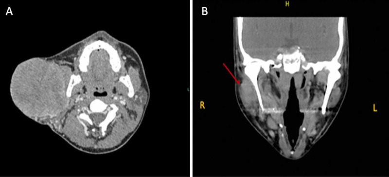
Figure 1. CT head and neck with contrast.
(A) First case: CT head and neck with contrast showing right parotid gland mass measuring 8.5 × 8.4 × 8.3 cm with central necrosis and mass effect on sternocleidomastoid muscle. (B) Second case: neck CT scan showing poorly demarcated mass (arrow) occupying superficial lobe of right parotid gland.

