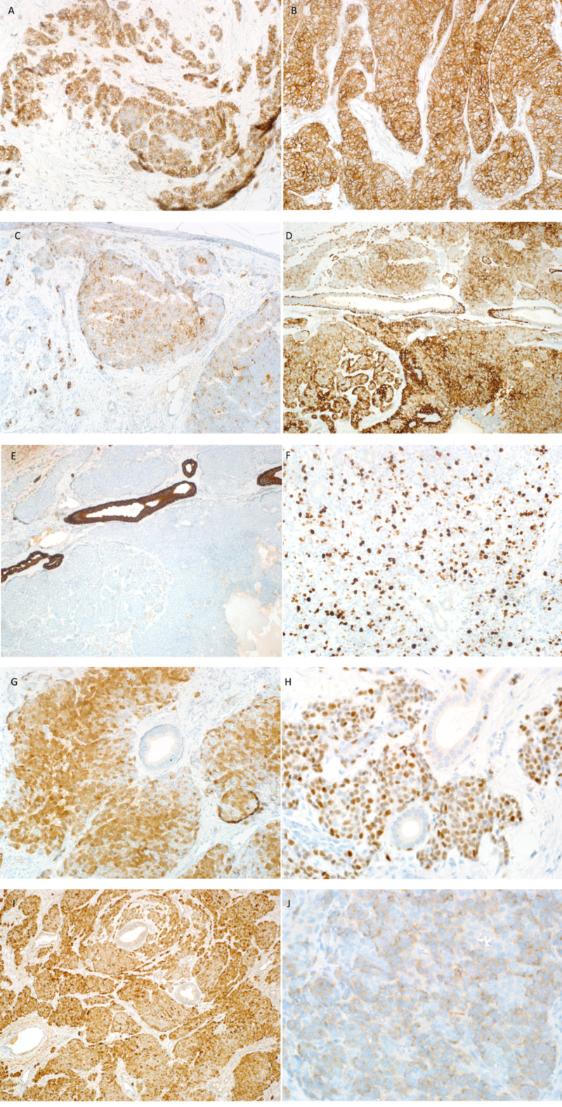
Figure 3. IHC stains of the first case.
(A) CD56: focally positive. (B) CD99: strong and diffuse membranous positivity in tumor cells. (C) Chromogranin: focally stains positive. (D) CK5/6: highlights myoepithelium of normal duct and stains tumor cells. (E) CK7: negative staining in tumor cells and positive in normal salivary duct. (F) Ki67: positive in more than 20%. (G) P16: nuclear and cytoplasmic positive staining in more than 75% of tumor cells. (H) P63: diffuse positive staining in tumor cells surrounding normal ducts. Some positive staining is also seen in myoepithelial cells. (I) PGP9.5: positive. (J) Synaptophysin: weakly positive. IHC, immunohistochemistry.

