. 2020 Dec 24;9:e64107. doi: 10.7554/eLife.64107

Figure 5. Incomplete elastic recoil of cleaved titin to the Z-disk.

(A) Nanogold immunoelectron micrographs of skeletal fibers labeled with different antibodies to I-band titin (T12, N2A, and proline-glutamate-valine-lysine-rich region (PEVK)). Shown are examples of Wt, Het, and Hom fibers after TEV protease treatment, held passively for 30 min at a stretched length. Red arrows point to Z-disks. (B) Recoil of elastic titin to the Z-disk in skeletal fibers quantified by measuring the antibody-epitope to Z-disk-center distance (n = 100 measurements/condition; SL range, 2.9–3.0 µm). (C) Passively fixed Hom TC cardiomyocytes labeled for the N2B titin element (central I-band) showing regular staining in controls (−GSN/−TEV; left). Titin cleavage by TEV protease (−GSN/+TEV, middle) or actin-removal by gelsolin treatment, followed by TEV treatment (+GSN/+TEV), caused partial (middle) or full (right) recoil of titin springs to Z-disks. (D) Quantification of titin recoil in Hom TC cardiomyocytes from images as in (C); measurement as in (B) (n = 200 measurements/group, evenly from 10 sarcomeres/group; SL range, 2.2–2.3 µm). Stats: ANOVAs with Tukey’s HSD post hoc procedure, further confirmed via ranked sum assessment (analysis not shown). Note: error bars are very small.

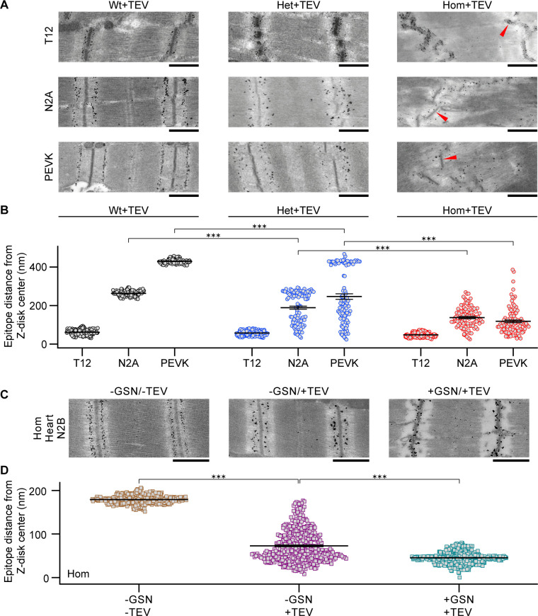
Figure 5—source data 1. Data and Stats for Figure 5.

Figure 5.

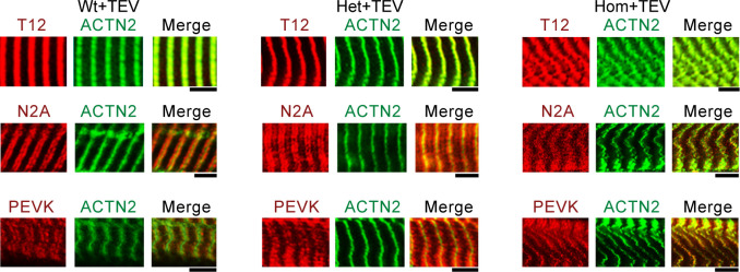
Figure 5—figure supplement 1. Immunofluorescence (IF) micrographs of skeletal fibers labeled with different antibodies to I-band titin (T12, N2A, and PEVK, red IF staining), and Z-disk marker α-actinin (ACTN2, green IF staining).

Figure 5—figure supplement 1.

Shown are examples of Wt, Het, and Hom fibers after TEV-protease treatment, held passively for 30 min at a stretched length. Scale bars, 5 μm.
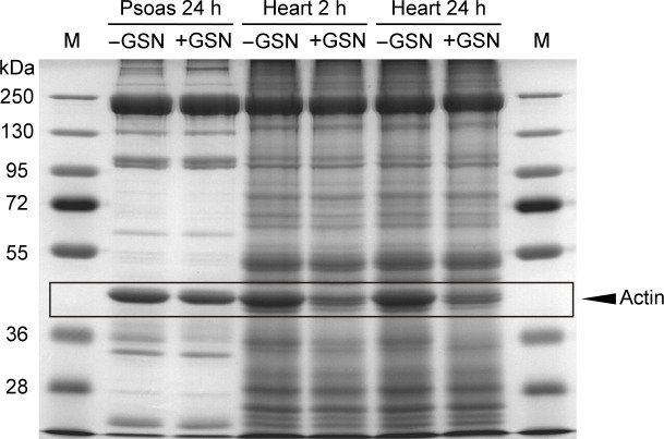
Figure 5—figure supplement 2. Coomassie-stained protein gel of skeletal (psoas) and cardiac muscle tissue.

Figure 5—figure supplement 2.

Skeletal muscle tissue was treated for 24 hr with Ca2+-independent gelsolin fragment (GSN), while cardiac tissue was treated for 2 or 24 hr. While cardiac actin is degraded by gelsolin, skeletal actin is not, as reported (Linke et al., 1997). Note that actin in the Z-disk is not degraded by the gelsolin fragment and can account for any remaining actin in cardiac tissue. These findings are supported by cardiac EM images, where gelsolin treatment leaves no visible thin filaments in the I-band (Figure 5C). M, marker.
Figure 5—figure supplement 3. Representative images of passively fixed, gelsolin-treated but not TEV-treated (+GSN/−TEV), Hom TC cardiomyocytes examined by transmission electron microscopy (EM) (left) or immuno-EM using antibodies to the N2B element of cardiac titin and nanogold-conjugated secondary antibodies (right).

Figure 5—figure supplement 3.

With thin filaments removed, I-band titin filaments can be observed with the characteristic higher density at the N2-line (red arrow). Thus, titin filaments are still intact after gelsolin treatment, suggesting that TEV-protease treatment is the cause of titin recoil toward the Z-disk (Figure 5C). Scale bar, 1 μm.