Figure 5.

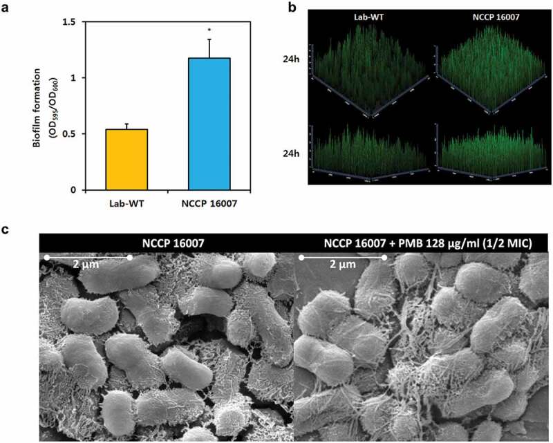
Biofilm formation of the NCCP 16007 strain. (a) Crystal violet quantification of biofilm formation in the Lab-WT and NCCP 16007 strains. (b) Confocal laser scanning microscopy (CLSM) images of biofilms in the Lab-WT and NCCP 16007 strains. Microscopic analysis was conducted using the Film TracerTM Sypro Ruby dye at 24 h. (c) SEM images of the NCCP 16007 strain. Pili-like biofilm structures were observed in the PMB-treated and non-treated samples. Bacterial cell samples were fixed, washed, dehydrated, dried, and coated with platinum prior to imaging. Data represent the mean (± standard deviation, SD; N = 3 biological replicates). *, P < 0.05 by Student’s t-test compared to theoretical value
