
Figure 1.
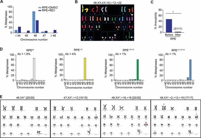
Aneuploid cells can survive in vitro. A Chromosome counts from metaphase spreads of RPE cells 20 h after treatment with reversine (REV) or DMSO vehicle-only control (n = 50; shown is the average of two independent replicates). B Representative spectral karyotype of an aneuploid RPE cell after reversine treatment. White arrow: t(X:10) present in the background (Fig EV1A-D). White arrowheads: chromosome gains after reversine treatment. C Percentage of aneuploid cells detected before and after single-cell clonal amplification; clones were classified aneuploid if their modal chromosome number calculated from 20 metaphase spreads ≠ 46 (*p < 0.05 by Fisher’s exact test). Polyploid cells with chromosome number >65 were seldom found. D,E Chromosome counts (d) and G-banding karyotypes (e) of a diploid (RPE46) and three aneuploid lines recovered after single-cell clonal amplification of reversine-treated RPE-1 cells. (D) Histograms represent the distribution of chromosome number per cell from metaphase spreads. Percentage of cells with structurally aberrant chromosomes (Ab) is shown top left; n = 50 metaphase spreads; shown is the average of two independent replicates. (E) Gained chromosomes and the t(X:10) translocation are indicated with red arrows; karyotypes (with derivative X chromosomes labeled with asterisks) are indicated at the top of the panels; karyotypic frequency is indicated in the brackets