
Figure 1.
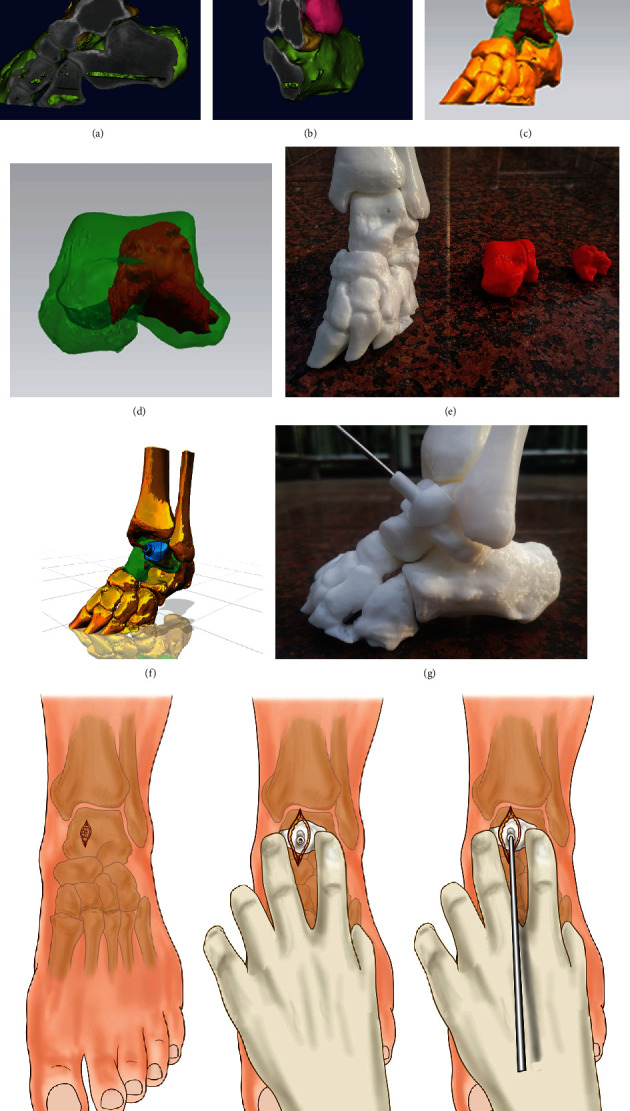
Preoperative preparation of 3D-printed lesion models and individualized guide system. (a, b) Talar lesions established based on the 3D CT images before surgery; (c, d) the software was used to distinguish lesions from healthy tissues; (e) 3D-printed lesion model that could be used for surgical planning and rehearsal; (f, g) design and preparation of individualized guides to assist in determining the position of the Kirschner wire. (h) The location of the lesion was entered through the incision, and the bony structure was confirmed; (i) the anatomical position was determined through the bony structure, and the cyst position was located with the guide; (j) the guide was used to assist the accurate positioning of the Kirschner wire.