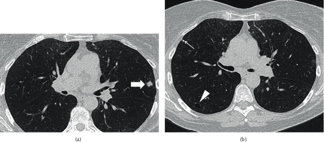
Figure 1.

(a, b) Axial chest CT images show an irregular shape, 1.1 cm, left upper lobe nodule with spiculated margins (block arrow), and a thin-walled parenchymal cyst in right lower lobe (arrowhead). Please note mild centrilobular, paraseptal emphysema, and a tiny, calcified right upper lobe nodule (thin arrow).
