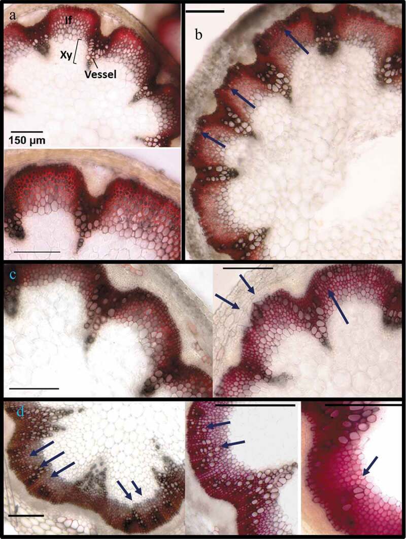
Figure 4.

Ectopic vessel development in stems of 8-week-old Arabidopsis plants over-expressing different phosphovariants of MYB75. Stems from 8-week-old plants were harvested and the bottom 5 cm of each stem was hand sectioned and stained in phloroglucinol-HCl. (a) Stem sections from Col-0 WT plants display normal distribution of vessels, within vascular bundles, where xylem vessels can be seen, as large perforated cells. In phloroglucinol-HCl stained sections, vessel walls appear dark reddish-brown, while interfascicular fibers are bright red/fuchsia arcs between the vascular bundles. (b) 35Spr:MYB75WT lines displayed relatively normal stem tissue organization, with a few large cells resembling vessel elements at the periphery of the interfascicular arc. (c) In 35Spr:MYB75T131A plants, large isolated cells or short cell files, resembling vessels can be seen in the interfascicular region. (d) In 35Spr:MYB75T131E plants prominent clusters of vessels could be seen in the interfascicular region
