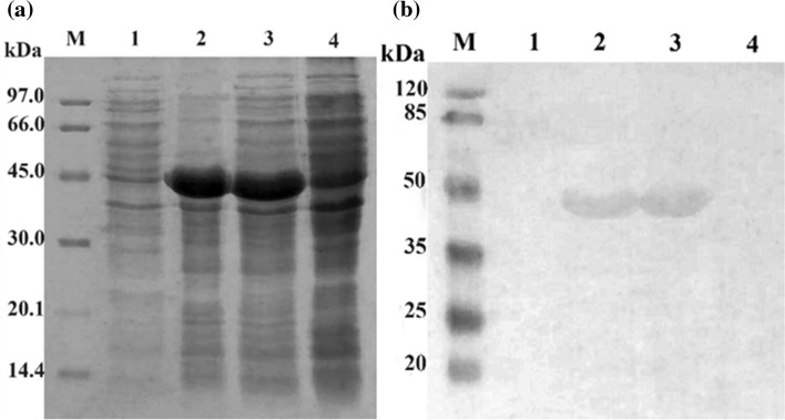
Fig. 3.
Coomassie brilliant blue staining of expressed SCARB2 analyzed by SDS-PAGE on 12.5% gel a and confirmed by Western blot probed with anti-His, b M, protein maker a and pre-stained protein marker b; 1–3, E. coli BL21 (DE3)/pET22b-SCARB2 (+ IPTG); 1, soluble phase; 2, insoluble phase; 3, total phase; 4, E. coli BL21 (DE3)/pET22b-SCARB2 (-IPTG)

