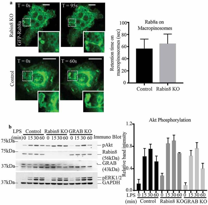
Figure 5.

Similar to GRAB, absence of Rabin8 does not affect Rab8a localisation and TLR signalling. (a) Live cell confocal spinning disc imaging of LPS-treated control and Rabin8 KO cells transiently overexpressing GFP-Rab8a showing recruitment to macropinosomes and tubules. Quantification of Rab8a retention on macropinosomes was measured by the total number of timeframes Rab8a is spent enriched on each macropinosome, and 5 cells of each cell line was used for quantification (n = 5). Scale bars, 10 µm. (b) Immunoblotting for levels of phosphorylated Akt in control, Rabin8 KO and GRAB KO cells treated with LPS over a 60 min time course. Quantification of Akt phosphorylation was performed by using the densitometric ratio between the band intensities of phosphorylated Akt and GAPDH. The immunoblot is representative and significance was measured via two-way analysis of variance (ANOVA) (n = 3)
