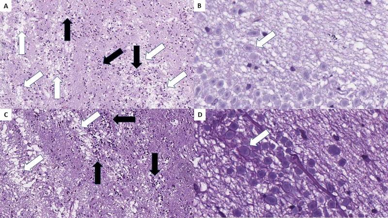
Figure 3. Necrotizing olfactory bulbitis as observed in both cases.
A and C: severe edema (white arrows) and diffuse inflammatory cell infiltration (black arrows), H&E stain, original magnification 100x; B and D: diffuse degenerative changes, H&E stain, original magnification 400x.
H&E: hematoxylin and eosin.

