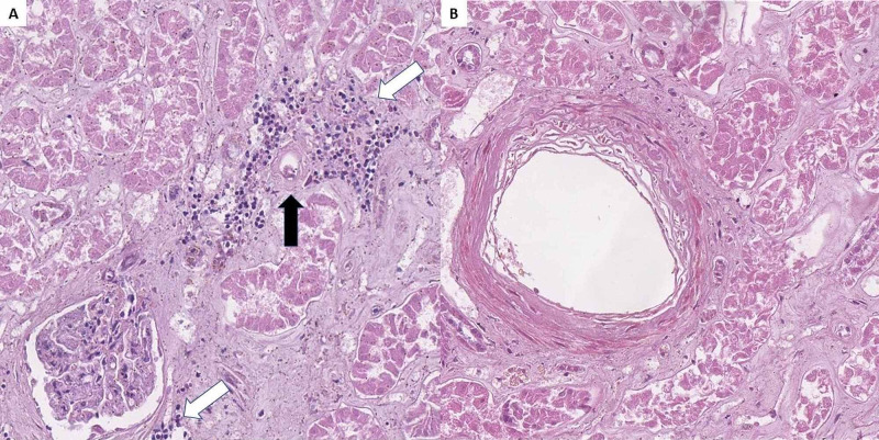
Figure 5. Renal histopathology.
A: lymphoplasmacytic inflammatory infiltration (white arrows) surrounding a small blood vessel with fibrinoid necrosis (black arrow), H&E stain, original magnification 200x; B: medium-sized blood vessel with fibrinoid necrosis, H&E stain, original magnification 200x.
H&E: hematoxylin and eosin.

