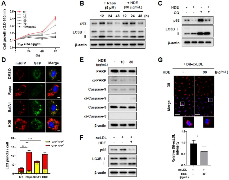
Fig. 2.
HDE inhibited cell proliferation and induced autophagy in HUVECs. (A) MTT assay was performed to analyze the effect of HDE on cell proliferation. HDE decreased cell proliferation in HUVECs in a concentration-dependent manner (0-100 µg/µl) for 72 h. (B) Autophagy was monitored on western blots of protein lysates of HUVECs following 30 µg/ml HDE treatments at 12, 24, and 48 h. (C) Cells were pretreated with chloroquine (25 µM) (or were not pretreated) for 1 h, before incubation in the presence or absence of 30 µg/ml HDE for 48 h. (D) HUVECs were transiently transfected with GFP-mRFP-LC3 and treated with rapamycin (10 µM) for 12 h, BafA1 (10 nM) for 3 h, or HDE (30 µg/ml) for 48 h, and then examined for changes in green and red fluorescence using a confocal microscope (Scale bar: 10 µm). The numbers of red puncta (GFP−RFP+) versus yellow puncta (GFP+RFP+) per cell in each condition were quantified. (E) Effects of HDE on apoptosis at 24 h. (F,G) HUVECs were incubated with 50 µg/ml oxLDL or DiI-labeled oxLDL in the absence or presence of 30 µg/ml HDE. After 48h, impaired autophagy was rescued by HDE and the amount of oxLDL contents was quan-tified by DiI intensity (scale bar: 20 µm). Results are presented as mean ± SEM values. Statistical significance was assessed using Student’s t-tests. ***P < 0.001; **P < 0.01.

