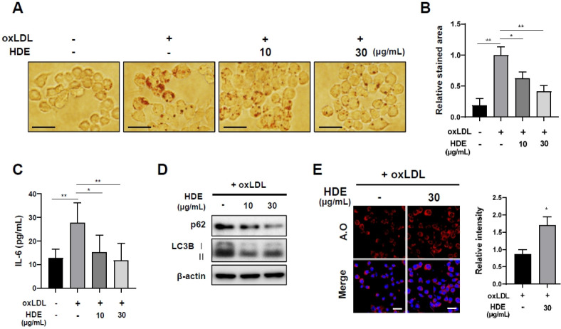
Fig. 3.
HDE reduced formation of foam cells by regulating autophagy flux. (A) Representative images of Oil Red O staining of RAW264.7 cells treated with HDE along with oxLDL (50 µg/ml) for 48 h (scale bar: 20 µm). (B) The area of stained lipid droplets in each group were quantified using Image J. (C) HDE inhibited IL-6 production in oxLDL-induced RAW 264.7 in a concentration-dependent manner. Under the same conditions, (D) autophagy markers were monitored on western blots. (E) Representative images of acridine orange staining results. RAW264.7 macrophages were co-treated with oxLDL (50 µg/ml) and HDE (30 µg/ml) for 48 h. Acidic vesicular organelles were detected (scale bar: 20 µm). Results are presented as mean ± SEM values. Statistical significance was assessed using Student’s t-tests. ***P < 0.001; **P < 0.01; *P < 0.05.

