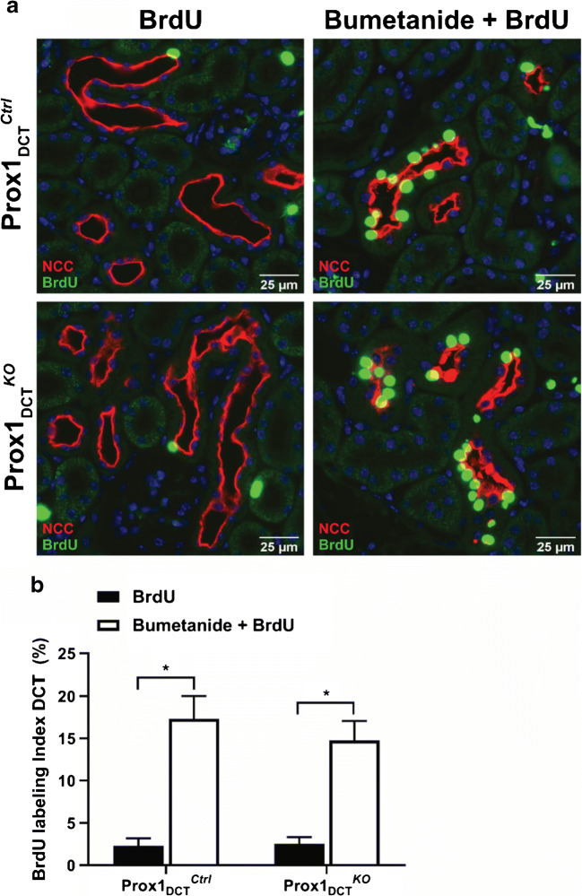
Fig. 4.
DCT cell proliferation rate in Prox-1DCTCtrl versus Prox-1DCTKO mice in response to a 3-day treatment with bumetanide. A Paraffin sections of Prox-1DCTCtrl mice kidneys (upper panel) and Prox-1DCTKO mice kidneys (lower panel) after a 3-day treatment with bumetanide and BrdU (right) or BrdU only (left). A double-immunostaining for NCC (red) and BrdU (green) allowed for the identification of DCT segments and proliferating DCT cells. B A significant increase in DCT cell proliferation rate was achieved in Prox-1DCTCtrl (p = 0.0001) and Prox-1DCTKO (p = 0.0007) by the administration of bumetanide and BrdU compared to the situation where mice received BrdU only. However, no difference in the proliferation rate between the different genotypes was observed. Data are mean ± SEM, n = 6 (BrdU only), n = 5 (Bumetanide + BrdU, ctrl), n = 4 (Bumetanide + BrdU, ko)

