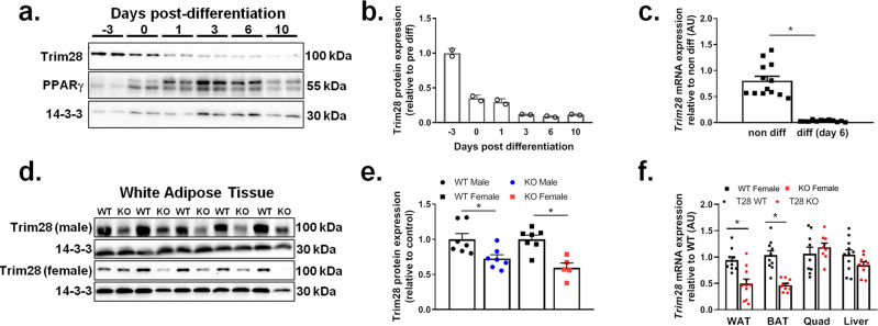
Fig. 1. Trim28 expression is reduced in differentiated adipocytes and in WAT from mice deleted for Trim28.
Protein and gene analyses were performed in 3T3-L1 adipocytes at different time points pre and post differentiation. a Representative immunoblot for Trim28, PPARγ, and 14-3-3 over a differentiation time course in 3T3-L1 cells, and (b) densitometry analysis of the Trim28 blots normalized to 14-3-3 and presented as relative to pre-differentiated 3T3-L1 Trim28 expression, n = 2. c Trim28 mRNA expression in non-differentiated (non-diff) and day 6 differentiated (diff day 6) 3T3-L1 cells, *p < 0.05 versus non-diff as analyzed by ANOVA with Fisher’s LSD post hoc testing, n = 13 non-diff, n = 12 diff. Cell culture experiments were repeated at least three times each. Protein and gene expression analyses performed in gonadal fat pads from Trim28 wild type (WT) and adipose-specific Trim28 KO (KO) mice. d Immunoblot of Trim28 and 14-3-3 in WT and adi-KO male and female fat pads. e Trim28 densitometry normalized to 14-3-3 relative to WT (n = 7 WT and adi-KO male mice, n = 7 WT and n = 5 adi-KO female mice), *p < 0.05 versus WT mice as analyzed by ANOVA with Fisher’s LSD post hoc testing. f Trim28 mRNA expression in WAT, BAT, quad, and liver of WT and adi-KO mice (n = 10 per group), *p < 0.05 versus WT mice as analyzed by ANOVA with Fisher’s LSD post hoc testing, n = 12 WT and n = 12 KO. All data are presented as mean ± SEM. WAT white adipose tissue, BAT brown adipose tissue, Quad quadriceps muscle.

