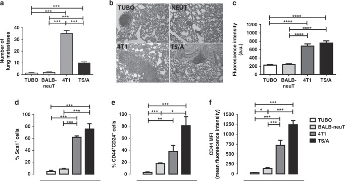
Fig. 3. 4T1 developed a higher number of lung metastases in vivo.
a Lung metastases count. ***P < 0.001. b Representative H&E staining of lung metastases of BALB/c mice s.c injected with TUBO, 4T1 or TS/A cells and BALB-neuT mice. Amplification ×10. c GLUT1 fluorescence intensity calculated for BALB-neuT, 4T1 and TS/A tumour slice compared to TUBO intensity. ****P < 0.0001. d–f Cytofluorimetric analysis of 10 mm mean diameter tumours (400 mm3) explanted from BALB-neuT mice or from BALB/c s.c. injected with TUBO, 4T1 or TS/A cells. Graphs showing the mean ± SD of the percentage of Sca1+ or CD44+CD24− cells and CD44 mean fluorescence intensity (MFI) from three independent experiments. *P < 0.05; **P < 0.01; ***P < 0.001.

