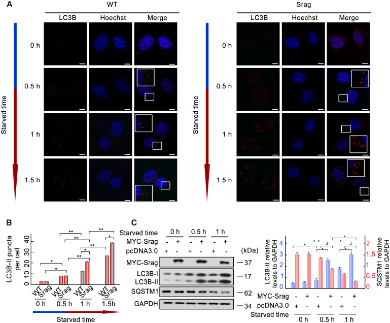
FIG. 4.
Srag overexpression promotes autophagy upon starvation induction. (A) Srag overexpression upregulated number of LC3B-II puncta. LC3B-II puncta were detected in wild-type or Srag overexpression HeLa cells cultured in the HBSS (Hanks balanced salt solution) medium for 0, 0.5, 1, and 1.5 h, respectively. Immunofluorescence was analyzed with anti-LC3B antibody and followed by confocal microscopy. LC3B-II puncta were detected in the cytoplasm by TRITC-conjugated ImmunoPure goat anti-rabbit IgG (red). The nuclei were revealed using Hoechst fluorescence. Enlarged boxes highlight LC3B-II signals. Scale bar: 5 μm. (B) Statistics of the LC3B-II puncta. The number of LC3B-II puncta was quantified from ∼20 cells for each group. The means ± SD are from three independent experiments. One-way ANOVA was performed. *P < 0.05; **P < 0.01. (n = 3 independent experiments). (C) srag overexpression upregulates LC3B-II level under starvation condition. HEK293T cells were transfected with equal amount of MYC-Srag or vector pcDNA3.0 (control) and cultured in the EBSS (Earle’s balanced salts solution) medium for 0, 0.5 and 1 h, respectively. Western blot analysis indicated that LC3B-II is upregulated by Srag overexpression in a time-dependent manner under starvation condition, whereas its downstream substrate SQSTM1 was downregulated. Cell lysates were analyzed by western blotting with the anti-LC3B and anti-MYC antibodies. GAPDH was used as an internal control. Western blots were quantified for LC3B-II/GAPDH and SQSTM1/GAPDH ratio. Data are presented as means ± SD. *P < 0.05; **P < 0.01 (n = 3 independent experiments).

