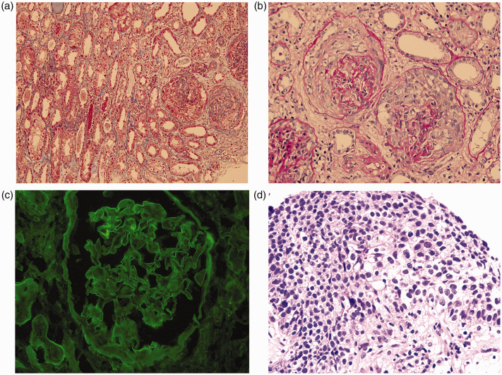
Figure 2.
Histopathology analysis revealed findings indicative of anti-GBM nephritis. (a and b) Renal biopsy showing diffuse cellular capsular crescent formation with focal necrosis (A: Periodic acid–Schiff stain, 200×, B: Masson's trichrome stain, 400×). (c) Diffuse linear deposition of IgG along the glomerular capillary basement membrane (immunofluorescence, 400×). (d) Squamous epithelial cells with severe dysplasia, enlarged and intensely stained nuclei, and obvious mitoses (Hematoxylin and eosin stain, 400×).
Abbreviation: GBM, glomerular basement membrane.

