
FIGURE 3.
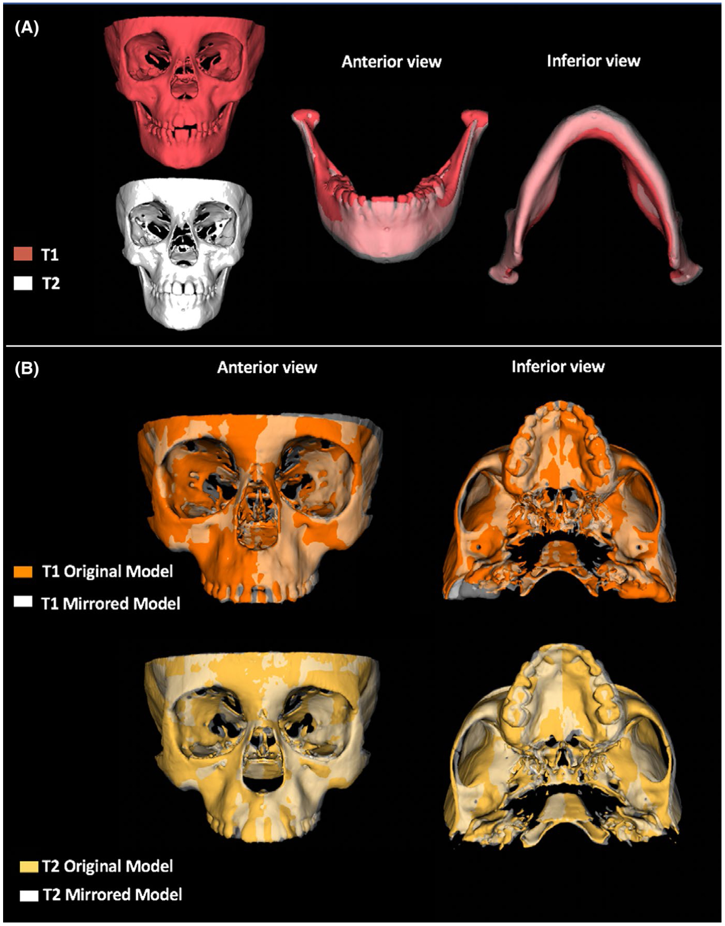
Summary illustration of the RME treatment results in asymmetry. A, 3D models in T1 and T2 of patient with functional unilateral posterior crossbite showing the shift change towards the left side after RME. B, 3D superimposition of patient with transverse maxillary deficiency and no posterior crossbite in T1 and T2 showing craniofacial regions more prominent at right side, as seen in zygomatic arch and maxilla