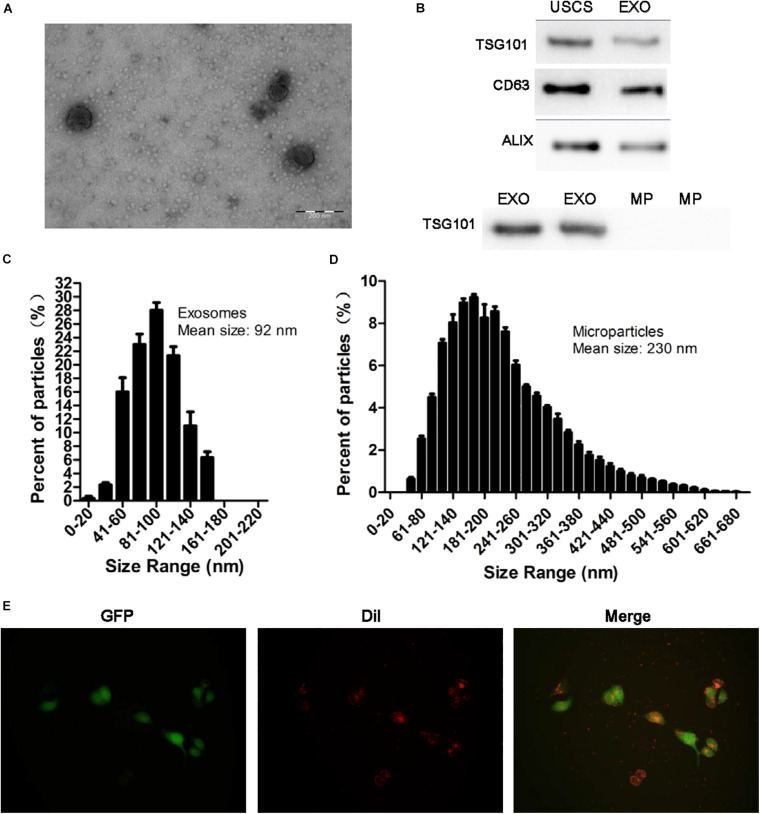
FIGURE 2.
Identification of exosomes and the uptake of exosomes from USCs by HK-2 cells. (A) Morphology of USC-Exos under TEM (scale bar = 200 nm). (B) The expression of CD63, TSG101, and ALIX in exosomes, MPs and USCs was determined by Western blot analysis. (C) The mean diameter and the concentration of exosomes were measured by Nanoparticle tracking analysis. (D) The mean diameter and the concentration of MPs were measured by Nanoparticle tracking analysis. (E) Green fluorescent protein (GFP) was introduced into HK-2 cells, exosomes labeled with red fluorescent dye Dil are shown to be up take by HK-2 cells.

