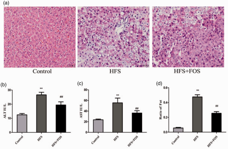
Figure 1.
FOS supplementation ameliorates HFS diet-induced histological and functional changes in the mouse liver. (a) H&E-stained liver tissue at 40× magnification. (b) Serum ALT levels (IU/L). (c) Serum AST levels (IU/L). (d) Ratio of fat in the liver. Data (b and c) are presented as mean ± standard deviation (n = 20 in each group). **P < 0.01 vs. the control group; ##P < 0.01 vs. the HFS group. FOS, fructo-oligosaccharides; H&E, hematoxylin and eosin; ALT, alanine transaminase; AST, aspartate aminotransferase; HFS, high-fat/high-sugar.

