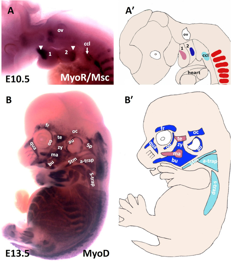
FIGURE 1.
Muscles of the mouse head and neck. (A) Lateral view of a stage E10.5 mouse embryo hybridized with a MSC (MyoR) probe. (A′) Schematic representation of E10.5 mouse embryo in the panel (A). In pink: first arch-derived muscle anlage. In blue: second arch-derived muscle anlage. In pale blue: cucullaris muscle anlage. MSC marked the myogenic core of the BA1 and BA2. Cucullaris muscle anlage is also labeled with MSC. (B) Lateral view of a stage E13.5 mouse embryo hybridized with a MyoD probe. (B′) Schematic representation of E13.5 mouse embryo in the panel (B). MyoD is expressed in all branchiomeric muscles and cucullaris muscle anlage. a-trap, acromio-trapezius; au, auricularis; BA1-2, branchial arches 1–2; bu, buccinator; ccl, cucullaris anlage; fr, frontalis; ma, masseter; oc, occipitalis; oo, orbicularis oculi; ov, otic vesicle; qua, quadratus labii; sp, splenius; stm, sternocleidomastoideus; s-trap, spino-trapezius; te, temporalis; zy, zygomaticus.

