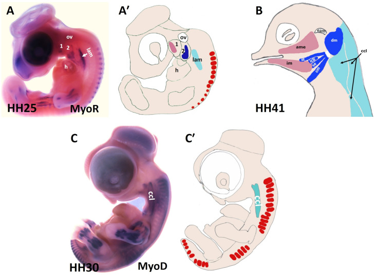
FIGURE 2.
Muscles of the chicken head and neck. (A,C) Lateral view of chicken embryos hybridized with DIG-probes for either MyoR (A) or MyoD (C). (A′,C′) Schematic representation of chicken embryos in the panels (A,C). (B) Lateral view of head muscles of a stage HH41 chicken embryo. The MyoD and MyoR delineated the BA1-derived muscles (pink), the BA2-derived muscles (blue) and the cucullaris muscle (pale blue). ame, adductor mandibulae externus; 1-2, branchial arches 1-2; ccl, cucullaris anlage; cm, caudal mylohyoideus; dm, depressor mandibulae; eam, external auditory meatus; h, heart; icb, interceratobranchialis; im, intermandibularis; lam, lateral plate mesoderm; ov, otic vesicle; se, serpihyoideus; st, stylohyoideus.

