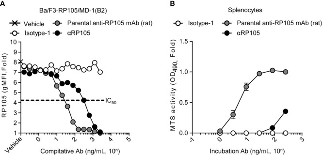
Figure 2.
αRP105 can also induce proliferation of splenocytes, but the activity is decreased. (A) Ba/F3 cells expressing RP105/MD-1 (B2) were incubated with the parental anti-RP105 mAb (rat), or the supernatant obtained from HEK293T cells expressing anti-HA (Isotype-1) or αRP105 after dilution from 2.5 × 10³ ng/ml, as indicated. Then, the cells were incubated with biotinylated parental anti-RP105 mAb (rat), followed by incubation with APC-conjugated streptavidin. Data were obtained using a BD LSRFortessa. Dose-dependent inhibitions are indicated as fold change normalized to geometric mean fluorescence (gMFI) from the incubation with 2.5 × 10³ ng/ml of parental anti-RP105 mAb (rat). The IC50 was determined by the average of the gMFI obtained from Isotype-1. (B) Splenocytes obtained from BALB/c mice were incubated with parental anti-RP105 mAb (rat), Isotype-1, or αRP105 after dilution from 2.5 × 10² ng/ml, as indicated, for 3 days. The proliferation and viability were assessed by an MTS assay. Data are indicated as the mean ± S.D. as fold change normalized to the absorbance at 490 nm from the incubation with 2.5 × 10² ng/ml of parental anti-RP105 mAb (rat). All indicated data are representative of at least two independent experiments.

