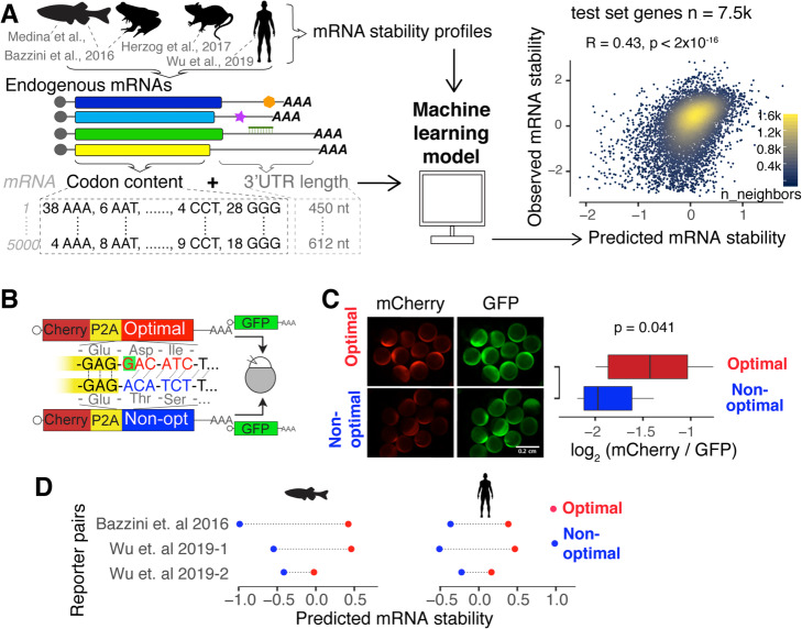
Fig. 1.
Codon composition predicts mRNA stability in vertebrates. a Scheme of the procedure to train a predictive model of mRNA stability. For each endogenous mRNA, the codon frequencies and the 3′UTR length are used as predictors to train a lasso regression model [28]. The scatter plot shows the point density of predicted and observed mRNA stability (test set genes n = 7576, Pearson correlation test). b Scheme of the 1 nucleotide out of frame reporters (optimal and non-optimal): two mRNAs that differ in the codon composition due to a single nucleotide deletion (G in red, highlight with green) which creates a frameshift. The encoding mCherry fluorescent protein was followed by a cis-acting hydrolase element (P2A) and then by a coding region enriched in optimal or non-optimal codons due to the frameshift. P2A causes ribosome skipping; therefore, the mCherry is not fused to the optimal or non-optimal encoded proteins. The mRNA reporter pairs were co-injected with mRNA encoding for GFP as an internal control [24]. c Fluorescence microscopy images of representative embryos at 8 h post-injection (hpi) with the indicated 1 nt out of frame reporter and GFP. Box plot displays fluorescence quantification at 8 hpi with each reporter. The mCherry fluorescence intensity was normalized to GFP intensity in each embryo (p = 0.041, paired t test). d mRNA stability predictions for 1 nucleotide out of frame reporters in fish and human cells. In all cases, the prediction for the optimal reporter is higher than that for the non-optimal (p = 0.007, binomial test)

