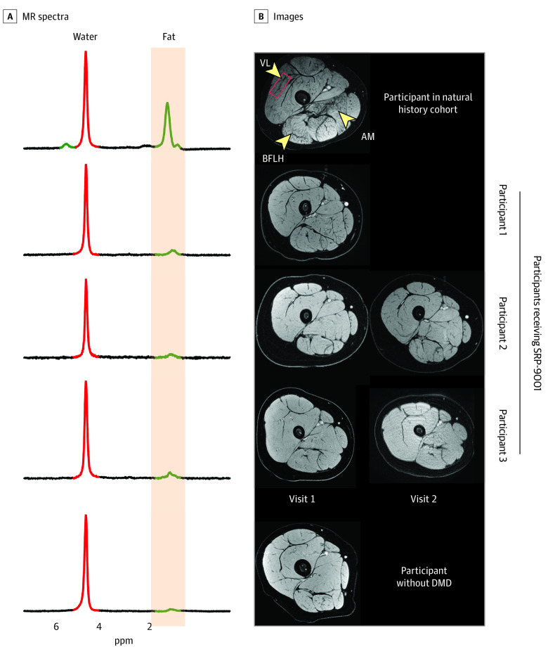
Figure 1. Example Magnetic Resonance (MR) Spectra and Images From Participants in the SRP-9001 Cohort, Natural History Cohort, and Control Cohort.
Spectra (A) and images (B) of representative participants from the natural history cohort, each participant who received SRP-9001 (participant 1, scan taken 18 months after treatment; participant 2, scans taken 6 and 18 months after treatment; participant 3, scans taken at 12 and 24 months after treatment), and a participant from the control cohort. AM indicates adductor magnus; BFLH, biceps femoris long head; DMD, Duchenne muscular dystrophy; and VL, vastus lateralis.

