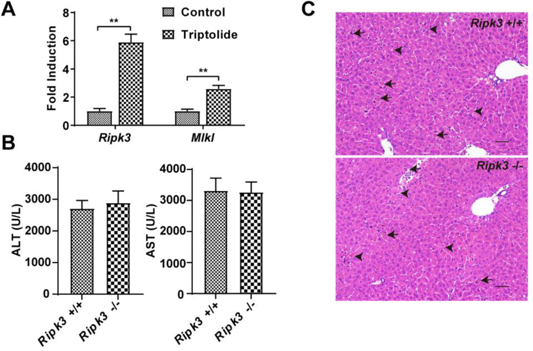
Figure 3.
Role of necrosis regulated by RIPK3 in triptolide-induced hepatotoxicity. (A) Expression of Ripk3 and Mlkl gene in control and mice treated with 0.8 mg/kg of triptolide. (B) Serum ALT and AST levels from control and 0.8 mg/kg triptolide-treated mice. (C) Micrographs of H&E stained liver sections (200×). Black arrows indicated apoptosis and black arrowheads indicated necrosis. The data were expressed as mean ± SEM (n=8). Statistical analyses were performed with Student’s t tests. *p <0.05, **p<0.01.

