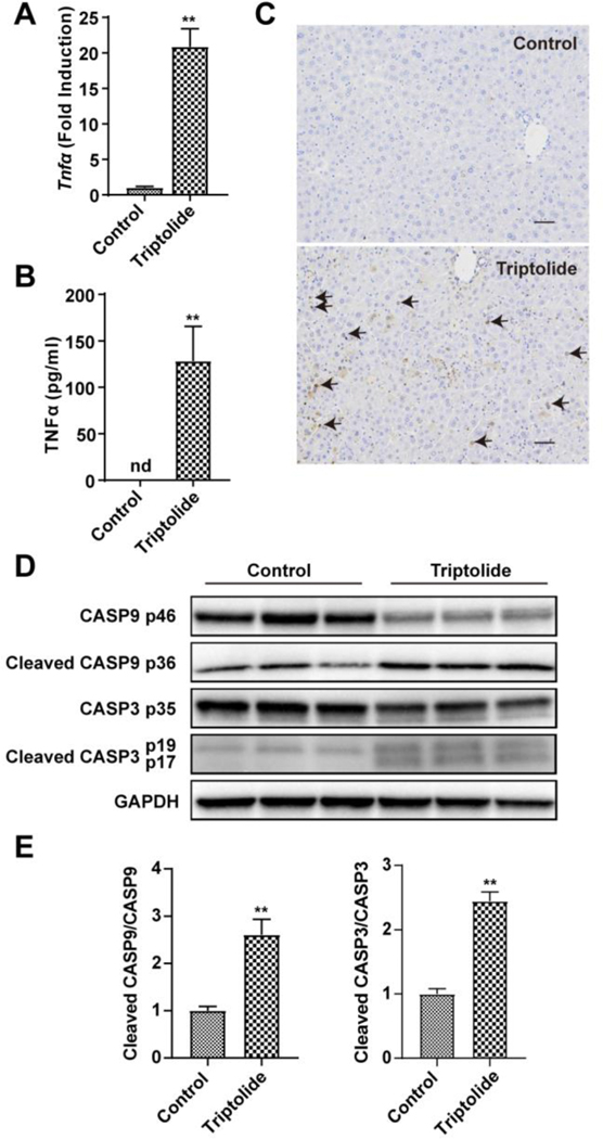
Figure 4.
Role of apoptosis in triptolide-induced hepatotoxicity. (A) QPCR measurement of the expression of Tnfα gene. (B) Plasma cytokine level of TNFα. (C) Micrographs of TUNEL assay in liver sections (200×). Black arrows indicated positive cells in TUNEL assay. (D) Western blotting analysis of expression of CASP9, cleaved CASP9, CASP3, cleaved CASP3 and GAPDH in the liver. (E) The ratio of cleaved form to full from of CASP9 and CASP3. The data were expressed as mean ± SEM (n=5–8). Statistical analyses were performed with Student’s t tests. **p<0.01.

