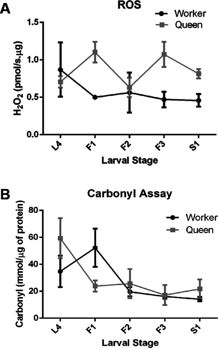
Figure 1. ROS levels and oxidative damage in fat body cells of honey bee queen and worker larvae. (A) H2O2 production rates were measured by the fluorometric Amplex® Red Hydrogen Peroxide/Peroxidase Assay in freshly dissected fat body tissue of the fourth (L4) and the first four substages of the fifth larval instar (F1-S1). Shown are means ± SEM (n= 3). (B) ROS-mediated damage to proteins was evaluated spectrophotometrically by the selective binding of 2,4-dinitrophenyl hydrazine (DNPH) to protein carbonyl groups in fat body homogenates of queen and worker larvae. Shown are means ± SEM (n=5). For statistical analysis results see text.

