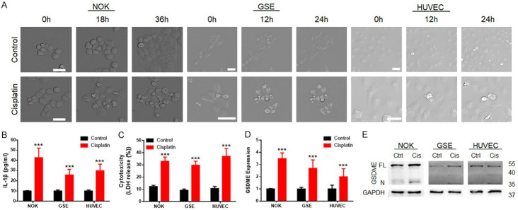
Figure 2.
GSDME participated in the regulation of pyroptosis in normal tissue cells during cisplatin-based chemotherapy. A. Microscope capture during the cell death process induced by cisplatin; the tested cells exhibited bubble-shaped death after cisplatin treatment (scale bar size: 50 μm). B. ELISA revealed elevated IL-1β expression in NOKs, GSEs and HUVECs; after cisplatin treatment; C. Cytotoxicity assay showing elevated LDH release in NOKs, GSEs and HUVECs after cisplatin treatment; D. PCR detection revealed that GSDME expression was upregulated in normal tissue cells after cisplatin treatment; E. Immunoblot showed the cleavage of GSDME (35 kDa) during cisplatin treatment of NOKs, GSEs and HUVECs; (cisplatin treatment: NOK 30 μM; GSE 20 μM; HUVEC 40 μM, 24 hours).

